Corpus: Skull


Synonym: cranium
1. Definition
2. Background
The number of bones attributed to the skull can vary in literature. Some authors include the hyoid bone and the auditory ossicles as part of the skull.
According to the Terminologia Anatomica, the hyoid bone is considered part of the skull. It is the only bone in the body not directly connected to another bone.
3. Anatomy
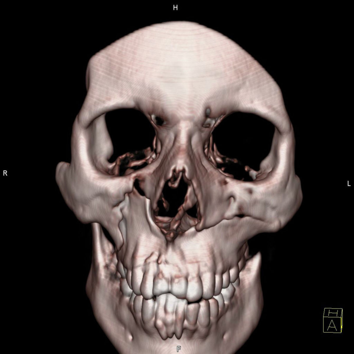
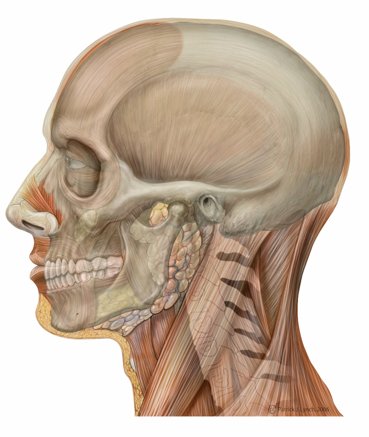
The skull is divided into the neurocranium and the viscerocranium. The transition between these two regions is marked at the base of the skull by the connection between the sphenoid and ethmoid bones. Skull shapes can also be categorized based on bony landmarks and measurements.
3.1. Neurocranium
The neurocranium is subdivided into the base of the skull (basis cranii) and the calvaria (skullcap). It consists of seven bones:
- Occipital bone
- Parietal bones (left and right)
- Frontal bone
- Temporal bones (left and right)
- Sphenoid bone
3.2. Viscerocranium
The viscerocranium forms the structural foundation of the face, shaping the orbits, nasal cavity, and oral cavity. It includes 15 bones:
- Ethmoid bone
- Nasal bones (left and right)
- Maxillae (left and right)
- Lacrimal bones (left and right)
- Zygomatic bones (left and right)
- Palatine bones (left and right)
- Inferior nasal conchae (left and right)
- Vomer
- Mandible
The boundary between the neurocranium and viscerocranium is best observed laterally, running from the upper edge of the orbit to the upper edge of the external auditory canal. The viscerocranium is therefore located ventral and caudal to the neurocranium.
4. Embryology
Cranial bones form through desmal ossification (direct bone formation) or chondral ossification (via a cartilage precursor). The viscerocranium predominantly arises from the desmocranium, while the neurocranium develops from both the desmocranium and chondrocranium.
5. Skull growth
In newborns, the cranial bones are not fully ossified, leaving gaps called fontanelles bridged by flexible connective tissue. The largest, the anterior fontanelle, is palpable in infants. These fontanelles close as the child grows, and the skull fully ossifies. In adults, the individual cranial bones remain distinguishable by their sutures.
6. Function
The skull's primary function is to protect the vital structures housed within it. These include components of the central nervous system, such as the brain, cerebellum, pons, and portions of the medulla oblongata, as well as essential sensory organs, including the eyes and the organs responsible for hearing and balance. Additionally, the skull facilitates food intake through the oral cavity and enables the grinding of food using the teeth.
Another significant function of the skull is thermoregulation, achieved through the diploic veins embedded within the cranial bones. These vessels play a crucial role in maintaining a stable temperature within the cranial cavity, ensuring optimal functioning of the structures it contains.
7. Deformities
The development of the skull during embryogenesis is a complex process that can be affected by errors, leading to deformities. Examples include:
- Anencephalus: This malformation involves the partial or complete absence of the cranial calvaria and the brain. It is classified as a neural tube defect caused by the failure of the anterior neuropore to close. During embryogenesis, the brain and neurocranium develop simultaneously; thus, if the brain does not form, the neurocranium is also not developed.
- Hydrocephalus: This condition typically arises from a defect in the ventricular system of the brain. It occurs when cerebrospinal fluid outflow is obstructed or when there is an imbalance between its production and resorption. The resulting accumulation of cerebrospinal fluid increases intracranial pressure, leading to the expansion of the neurocranium in areas that have not yet ossified.
8. Source
- Reith W: Hydrocephalus. Radiologist. 52: 805. 2012