Corpus: Frontal bone

1. Definition
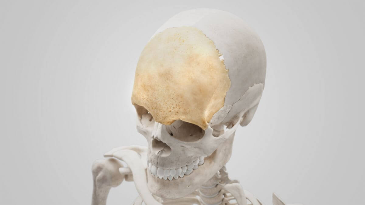
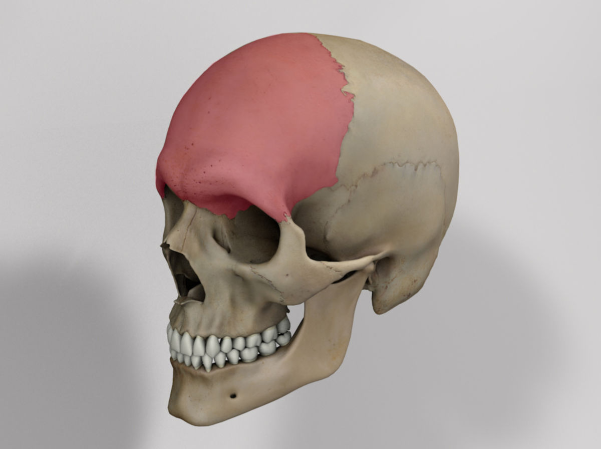
The frontal bone is a shell-shaped bone of the skull that forms the bony base of the upper orbital rims and the forehead. It is part of the neurocranium.
2. Anatomy
The frontal bone can be roughly divided into two parts:
- Frontal squama
- Orbital part
Some authors define a third part, the nasal part, which forms the middle section between the two orbital parts.
2.1. Frontal squama
The frontal squama forms the bony framework of the forehead. It consists of a convex external surface and a concave internal surface, each with characteristic structures.
2.1.1. External surface
The external surface is curved and shows the frontal suture (sutura frontalis or sutura metopica) in the midline, which divides the bone into two parts during childhood. The frontal eminences can be found on both sides, about 3 cm apart. This elevation, also known as the frontal tuberosity, varies greatly among individuals and is particularly prominent in children.
Below the eminence, separated by a shallow bone pit, are the two superciliary arches, which are connected in the center by the glabella. The arches are more pronounced in men than in women. Further down, the frontal squama ends in a curved bony edge, the supraorbital margin, which forms the upper edge of the orbit. It separates the frontal squama from the neighboring orbital part.
2.1.2. Internal surface
The internal surface is concave and features a vertical furrow in the upper part of its midline, the superior sagittal sinus groove, which receives the superior sagittal sinus. The falx cerebri attaches to the protruding edges of the sulcus. Further down, these bony ridges converge to form the frontal crest, which ends in a small depression called the foramen cecum.
On both sides of the sulcus, there are numerous bulges to accommodate cerebral convolutions and smaller grooves that house branches of the middle meningeal artery.
2.2. Orbital part
The orbital part forms the roof of the eye and nasal cavities. It consists of two relatively thin, almost triangular bone plates, known as the orbital plates, and the recess between them for the ethmoid bone, called the ethmoid notch.
3. Ossification
The frontal bone is formed by intramembranous ossification from three bone nuclei. A median frontal suture (sutura metopica) can occasionally be recognized between the originally paired frontal bones. It is a remnant of the frontal suture, which normally ossifies completely by the age of two to six years.
The granular foveolae are another remnant. These are thin-walled depressions in the bone caused by the pressure of the arachnoid granulations on the dura mater.