Corpus: Orbit


Synonym: orbital cavity; orbit; eye socket
1. Definition
The orbit is the cavity in the front region of the skull that houses the eye, along with its associated structures, blood vessels, and nerves.
2. Anatomy
2.1. Overview
The orbit is made up of parts of several skull bones. It has a roughly four-sided pyramid shape, with its base facing forward and its tip extending deep into the skull. It provides a bony protective enclosure for the eye. The bones contributing to the structure of the orbit include:
2.2. Boundaries
The roof of the orbit is formed in the front by the frontal bone and in the back by the sphenoid bone (lesser wing of the sphenoid).
The floor of the orbit is primarily made up of the orbital surface of the maxilla and the zygomatic bone. A small portion in the back of the floor is formed by the palatine bone. The lowest point of the orbital floor is often referred to as the orbital margin of the maxilla.
The lateral wall is formed by the zygomatic bone and the greater wing of the sphenoid bone.
The medial wall, which is very thin, is composed (from front to back) of the maxilla, lacrimal bone, ethmoid bone (lamina papyracea), frontal bone, and the lesser wing of the sphenoid bone.
2.3. Openings
The frontal entrance to the orbit, or the orbital opening, is called the aditus orbitalis. It is bordered by the bony orbital rim.
The orbit is connected to the middle cranial fossa through the superior orbital fissure and the optic canal. The inferior orbital fissure links the orbit to the pterygopalatine fossa. These openings allow important structures such as nerves and vessels to pass through.
The lacrimal bone and maxilla form the lacrimal sac fossa, which is bounded by the anterior and posterior lacrimal crests. This area contains the nasolacrimal duct, which drains tears into the nasal cavity.
The infraorbital groove leads into the infraorbital canal, providing a pathway for the infraorbital nerve and vessels.
The anterior and posterior ethmoidal foramina allow passage of the corresponding nerves and vessels between the orbit and the cranial cavity.
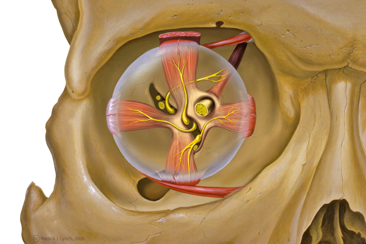
2.4. Contents
The orbit contains the following structures:
- Eyeball (bulbus oculi)
- Extraocular muscles (responsible for eye movement)
- Retrobulbar fat (cushioning fat tissue behind the eyeball)
- Lacrimal apparatus: including the lacrimal gland, lacrimal sac, and nasolacrimal duct
- Nerves: including the optic nerve (cranial nerve II), ophthalmic nerve (branch of cranial nerve V1), oculomotor nerve (cranial nerve III), trochlear nerve (cranial nerve IV), abducens nerve (cranial nerve VI), and short ciliary nerves
- Blood vessels: such as the ophthalmic artery and vein
- Ciliary ganglion (a parasympathetic ganglion involved in innervating the eye)
3. Histology
The orbit consists of the typical bony parts of the flat bones of the skull. The bony orbit is separated from the contents by a layer of periosteum, which is referred to as the periorbita.
4. Function
The orbit serves to hold, fix and protect the eye and as the origin of the eye muscles.
5. Clinic
The orbit is often involved in midface fractures. This can lead to a stepped formation of the orbital rim. The direct impact of force on the eye can rupture the orbital floor. This is referred to as a blow-out fracture.
The orbit and its contents are also affected in a number of autoimmune diseases. Typical examples of these so-called orbitopathies are myositis of the eye muscles and Graves' disease.