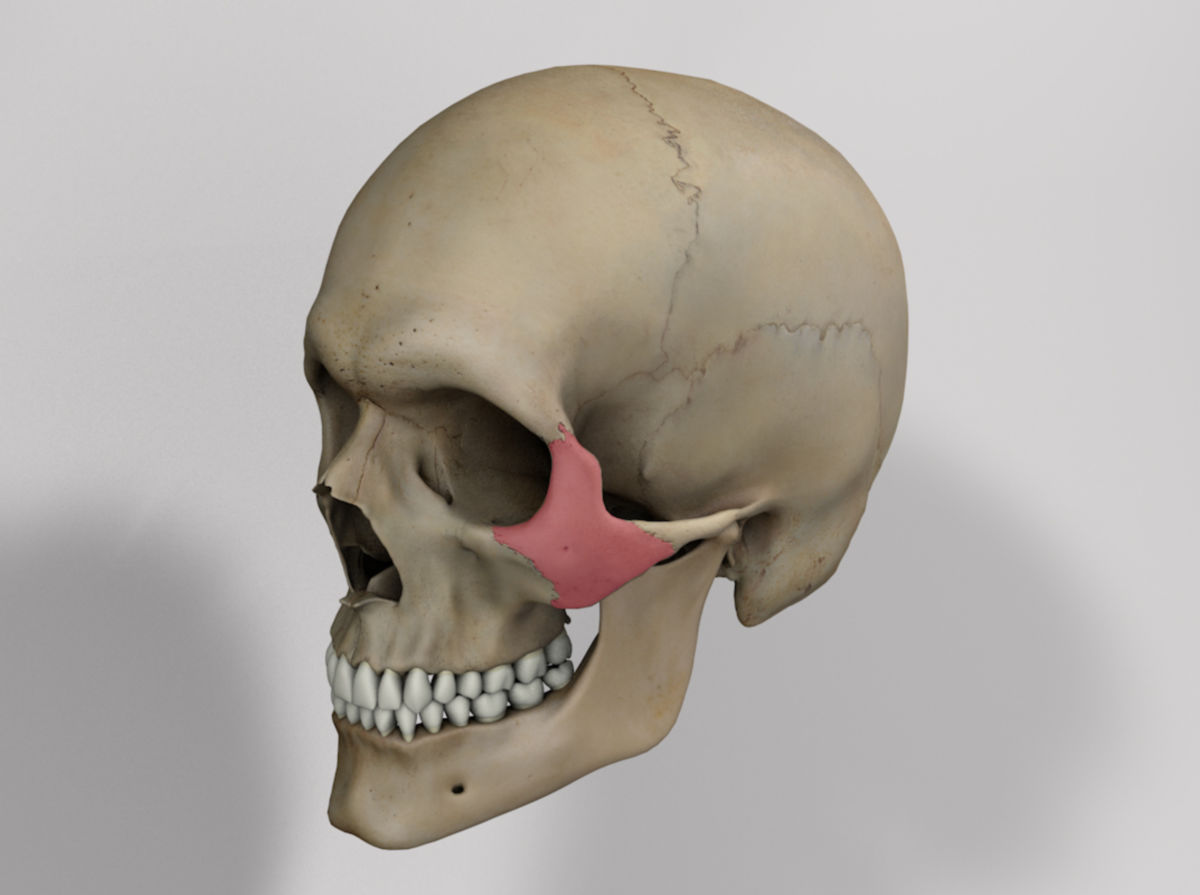
Corpus: Zygomatic bone


Synonym: malar bone
1. Definition
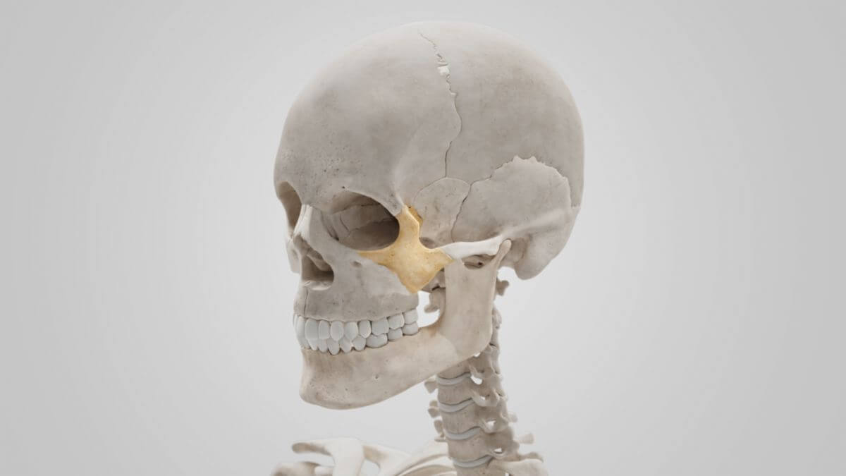
2. Anatomy
2.1. Surfaces
The zygomatic bone has three surfaces: the lateral surface, the temporal surface, and the orbital surface.
2.1.1. Lateral surface
The lateral surface, also known as the malar surface, is convex and features a bony opening in its center called the zygomaticofacial foramen. The zygomaticus major and zygomaticus minor muscles originate from this area. This surface is palpable as the "cheekbone" through the soft tissues of the face.
2.1.2. Temporal surface
The concave inner surface, the temporal surface, slopes dorsally and medially. It comprises the temporal fossa in the upper part and the infratemporal fossa in the lower part. The anterior part has a rough, almost triangular area of bone that articulates with the maxilla. The zygomaticotemporal foramen is located within the temporal surface.
2.1.3. Orbital surface
The orbital surface, also referred to as the orbital process by some authors, is smooth and forms part of the floor and lateral wall of the orbit, along with the maxilla and the sphenoid bone. The zygomaticoorbital foramen is located approximately in its center.
2.2. Processes
The cranially pointing frontal process articulates with the zygomatic process of the frontal bone. It forms the lateral orbital rim.
The maxillary process has a plump, triangular cross-section that articulates with the zygomatic process of the maxilla. It points forwards and medially. The origin of the levator labii superioris muscle is located at the orbital section of its anteroinferior margin.
The dorsally pointing temporal process articulates with the zygomatic process of the temporal bone, and together they comprise the zygomatic arch. The original surface of the masseter muscle lies on its lower edge.
3. Clinic
A zygomatic fracture can occur as a result of significant trauma, such as from an accident or physical altercation.