Corpus: Temporal bone


1. Definition
The temporal bone is a complex structure in the human skull, housing the middle and inner ear. It forms the socket of the temporomandibular joint.
2. Overview
The temporal bone houses the middle and inner ear and is involved in the temporomandibular joint. It consists of four parts:
- Temporal bone scale
- Tympanic part with auditory canal and middle ear
- Mastoid process with mastoid cells
- Petrous bone with inner ear
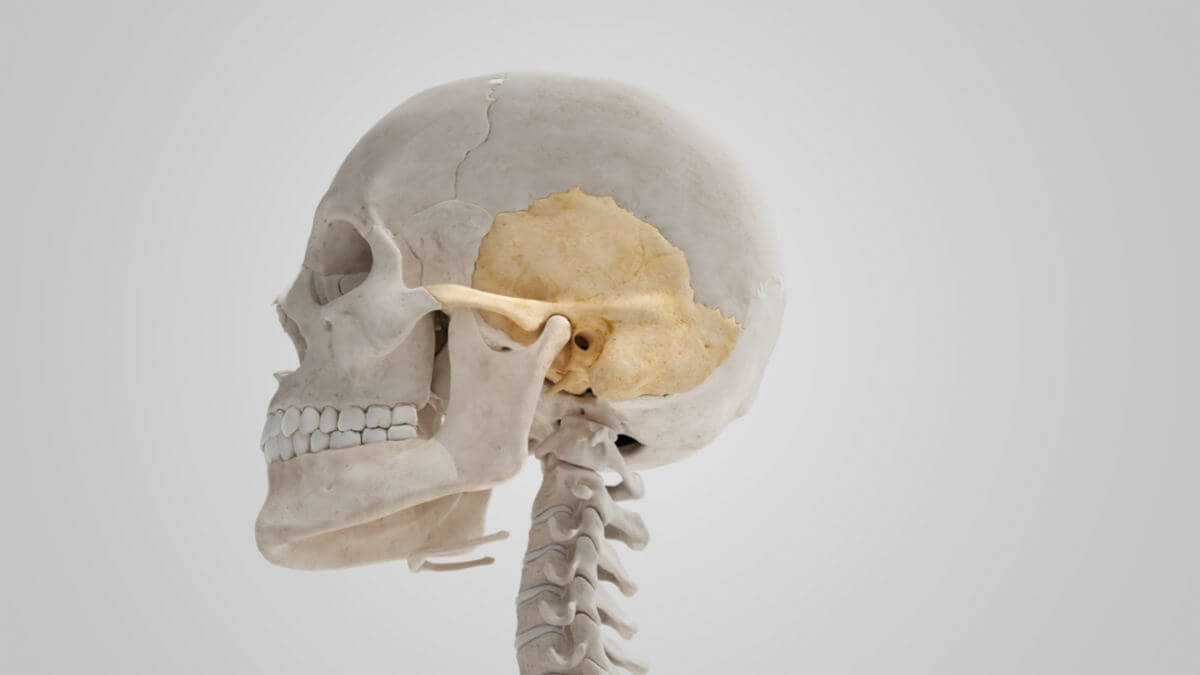
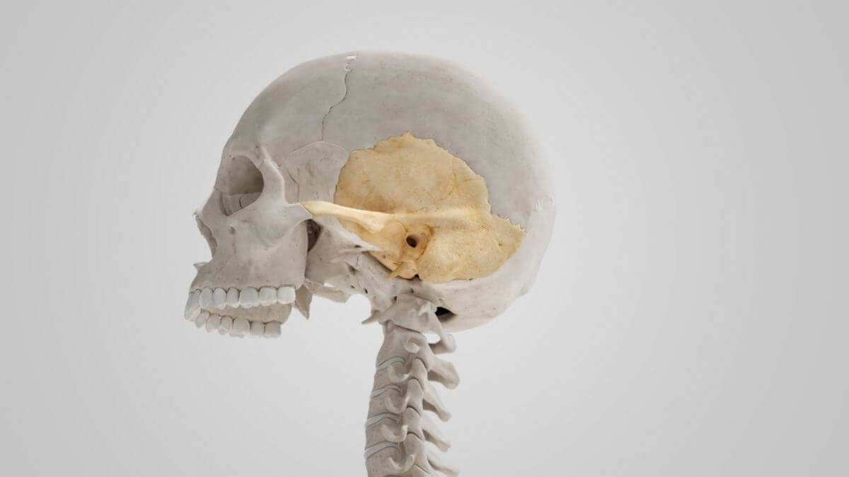
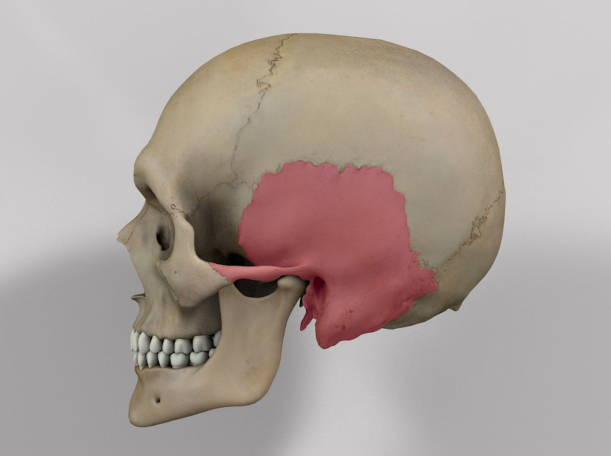
3. Temporal bone scale
3.1. Surfaces
3.1.1. Temporal surface
The temporal surface of the temporal bone scale is smooth and convex. It serves as the origin of the temporalis muscle and forms part of the temporal fossa. In the posterior part, a vertically ascending groove can be recognized, where the middle temporal artery runs.
A prominent bony process, the processus zygomaticus ossis temporalis, arises from the anterior part of the temporal surface. Its upper edge is thin and sharp, serving as an attachment for the temporal fascia, while the lower edge is thicker and curved, where the fibers of the masseter muscle originate. The lateral surface of the process borders on the subcutaneous tissue. Its anterior end articulates with the temporal process of the zygomatic bone, comprising the zygomatic arch. At the posterior end of the zygomatic process, two bony ridges connect to the temporal bone scale. The mandibular fossa, covered with cartilage, is also found here. The zygomatic process continues dorsally in a flat bony ridge, the supramastoid crest, which serves as an attachment for the temporal fascia and delimits the origin of the temporalis muscle. The boundary between the squamosal and the mastoidal parts runs approximately 1 cm below the groove, sometimes recognizable by the remains of a suture.
3.1.2. Cerebral surface
The cerebral surface of the temporal bone scale is concave. It shows shallow impressions from the temporal lobe of the brain and bony grooves for the branches of the middle meningeal artery.
3.2. Margins
The superior margin of the temporal bone is thin, with the external tabula overlapping the internal tabula. It articulates with the parietal bone at the squamosal suture. The anteroinferior margin is thick and serrated, articulating with the greater wing of the sphenoid bone.
4. Tympanic part
The tympanic part of the temporal bone is a curved bony structure located caudal to the pars squamosa. It lies in front of the mastoid process and surrounds the external acoustic meatus. The tympanic part develops as a separate bone that fuses with the other parts of the temporal bone as the skull grows.
4.1. Surfaces
The posterosuperior surface is concave, forming the anterior wall, the floor, and parts of the posterior wall of the external acoustic meatus. Medially, there is a small furrow where the eardrum attaches, known as the tympanic sulcus.
The anteroinferior surface is slightly concave, comprising the posterior boundary of the mandibular fossa and contacting the retromandibular part of the parotid gland.
4.2. Margins
The lateral edge of the tympanic part, where the cartilaginous part of the external auditory canal attaches, has a rough bone surface into which connective tissue fibers radiate. The posterior margin connects with the squamosal and the mastoid part, comprising the anterior boundary of the tympanomastoid fissure. The upper margin is in contact laterally with the back of the postglenoid process and medially with the petrotympanic fissure. The medial part of the lower margin is thin and tapers sharply, while the lateral part splits to form the vaginal process, surrounding the root of the styloid process. The styloid process and the stylohyoid ligament develop from the second branchial arch, the Reichert cartilage.
4.3. Other structures
The bony external acoustic meatus is about 2 cm long, extending inward and slightly rostrally. The sagittal section shows an oval or elliptical cross-section. The floor, as well as the anterior and lower part of the posterior wall, are formed by the tympanic part. The ceiling and the upper part of the posterior wall belong to the squamosal part.
5. Mastoid process
5.1. Surfaces
5.1.1. External surface
The outer surface of the mastoid part is rough, serving as the origin for the occipitalis muscle and the posterior auricularis muscle. It is penetrated by numerous foramina, with the larger mastoid foramen near the posterior margin. This foramen is traversed by a vein to the transverse sinus and a small branch of the occipital artery. The presence and location of this foramen are variable, sometimes located in the occipital bone or in the neighbouring occipitotemporal suture.
In the lower part, a prominent, conically tapering bony bulge, the mastoid process, is generally more pronounced in men than in women. This process serves as the insertion or origin of various neck muscles, particularly the sternocleidomastoid muscle, the splenius capitis muscle, and the posterior belly of the digastric muscle.
On the medial side of the mastoid process, a clear incision, the mastoid notch, is visible, marking the origin of the posterior belly of the digastric muscle. The occipital artery lies immediately medial to this in a small depression, the sulcus arteriae occipitalis.
5.1.2. Internal surface
The sigmoid sinus groove, a deep bony groove that receives the sigmoid sinus, can be seen on the inner surface of the mastoid part. The floor of the sulcus is separated from the pneumatisation spaces of the mastoid by a thin bone layer.
5.2. Margins
The superior margin of the mastoid part is broad and serrated, articulating with the mastoid angle of the parietal bone. The posterior margin articulates with the inferior margin of the occipital bone between its lateral angle and the jugular process. The anterior margin is fused with the descending bony process of the squamosal part. The lower edge of the mastoid part forms parts of the external acoustic meatus and the tympanic cavity.
5.3. Spaces
The mastoid part contains numerous pneumatisation spaces, known as mastoid cells, whose number, size, and extent vary among individuals. In the upper and frontal parts, these cells are large and irregularly shaped, containing air. Their size decreases caudally, sometimes appearing as small, narrow medullary cavities or being completely absent.
The anterosuperior region of the mastoid contains a large cavity, the tympanic antrum, which is separate from the mastoid cells but communicates with them via bone openings. The tympanic antrum contains air and is lined by extensions of the tympanic cavity mucosa. It opens rostrally into the epitympanic recess of the tympanic cavity and is separated from the middle cranial fossa by a thin bone plate, the tegmen tympani. On the medial wall, the lateral semicircular canal of the inner ear projects into the cavity.
6. Petrous bone
7. Topography
The temporal bone is adjacent to the following bones:
- Occipital bone (posterior)
- Parietal bone (superior)
- Zygomatic bone (anterior)
- Sphenoid bone (medial)