Corpus: Parietal bone


1. Definition
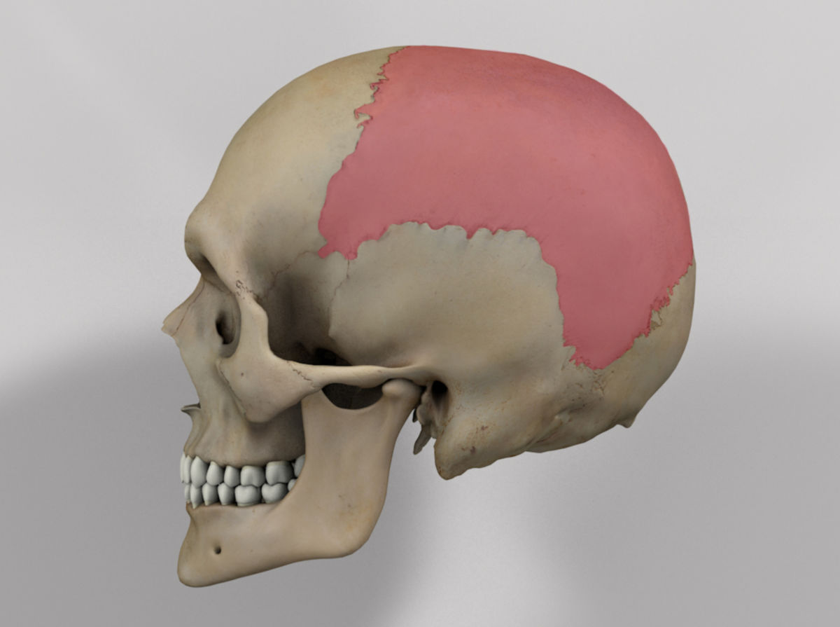
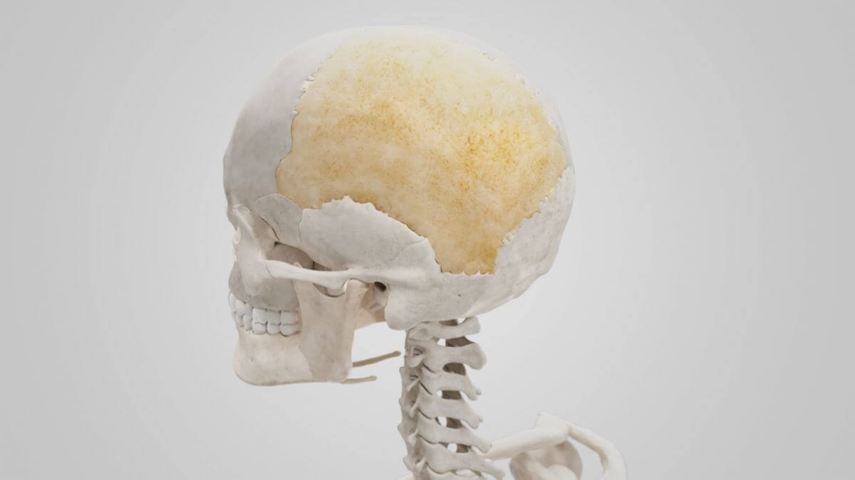
The paired parietal bone is a skull bone that belongs to the neurocranium. It forms the roof of the skull and most of the lateral wall of the skull.
2. Surfaces
2.1. External surface
The outer surface of the parietal bone is convex and smooth. Approximately in its center, it has a bulbous bony elevation called the parietal tuberosity, which represents the area where ossification of the parietal bone begins. Inferior to the tuberosity, two curved, parallel bone ridges, the superior temporal line and the inferior temporal line, cross over the bone surface. The superior temporal line serves as the attachment of the temporal fascia, while the inferior temporal line marks the upper end of the origin of the temporalis muscle.
2.2. Internal surface
The inner side of the parietal bone is concave. Flat depressions correspond to the adjacent cerebral convolutions (gyri). Branching furrows extend from inferior to superior, take the courses of the branches of the middle meningeal artery, and are called groove for middle meningeal artery.
A shallow bony groove at the upper edge, supplemented by the corresponding structure of the opposite parietal bone, forms the sagittal sulcus for the superior sagittal sinus. The falx cerebri attaches to the edge of this sulcus. Near the sulcus, especially in older individuals, depressions for the arachnoid granulations are recognizable as granular foveolae. A small parietal foramen also occurs inconsistently at the upper edge of the parietal bone.
3. Margins
3.1. Sagittal margin
The sagittal margin of the parietal bone is the longest and thickest. It is serrated and articulates with the bone at the opposite side. The suture between the two bones is the sagittal suture.
3.2. Frontal margin
The frontal margin is deeply serrated and articulates with the frontal bone. The junction of the two bones comprises the coronal suture. The right and left parietal bones each contribute 50 % to this suture. The point where the sagittal suture meets the coronal suture in a T-shape in the median plane is called the bregma.
3.3. Squamous margin
The squamous margin can be divided into three parts:
- The anterior part articulates with the greater wing of the sphenoid bone. It is thin and pointed. The inner plate overlaps the outer plate and is thus overlapped by the tip of the sphenoid wing.
- The middle part is concavely indented and is overlapped by the squamous part of the temporal bone.
- The posterior part is thick and serrated, articulating with the mastoid part of the temporal bone.
3.4. Occipital margin
The occipital margin is deeply serrated and forms the lambdoid suture during articulation with the occipital bone. The point where the sagittal suture meets the lambdoid suture is called the lambda.