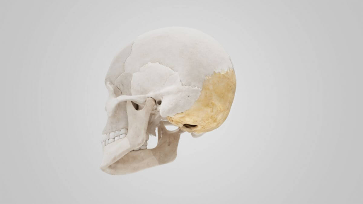
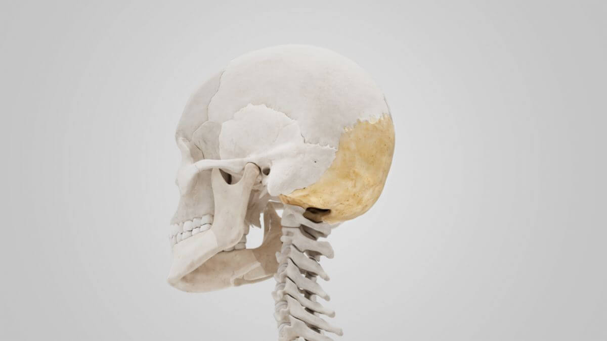
Corpus: Occipital bone


from Latin: occiput - back of the head
1. Definition
The occipital bone is a flat cranial bone and part of the neurocranium. It borders the parietal bones above via the lambdoid suture, the temporal bones laterally, and the sphenoid bone below.
2. Anatomy
The occipital bone is located in the posterior lower part of the skull. It is a trapezoidal bone of the neurocranium and, together with the temporal bones, forms the posterior cranial fossa.
The occipital bone can be subdivided into four parts:
- Basilar part: The unpaired thick, roughly quadrangular part rostral to the foramen magnum, forming the posterior part of the base of the skull.
- Lateral parts: The two paired parts on both sides lateral to the foramen magnum.
- Squamous part: The unpaired flat, bowl-shaped part dorsal to the foramen magnum.
2.1. Basilar part
The basilar part runs rostrally from the foramen magnum and comprises the clivus of the occipital bone in the posterior fossa. The outer aspect features the pharyngeal tubercle, serving as the attachment point for the pharyngeal raphe. Attachment points for the longus capitis and rectus capitis anterior muscles are also located on this outer aspect.
2.2. Lateral parts
The lateral parts contain the occipital condyles, located on the outer side lateral to the foramen magnum. Together with the atlas, they comprise the atlanto-occipital joint. The occipital condyles are each penetrated by the hypoglossal canal. Posterior to the occipital condyles is the condylar fossa, which in some individuals is perforated by the condylar canal. Lateral to the occipital condyles is the jugular process, forming the posterior part of the jugular foramen. The rectus capitis lateralis muscle attaches to the lateral part of the occipital bone. The inner side of the lateral part features the jugular tubercle, which covers the hypoglossal canal.
2.3. Squamosa part
On the outer surface, the four nuchal lines and the external occipital protuberance, a point of origin for the descending part of the trapezius muscle, can be recognized. On the inside, the internal occipital protuberance, the transverse sinus sulcus, and its continuation into the sigmoid sinus sulcus are visible.
2.4. Foramen magnum
Posterior to the basilar part, the foramen magnum comprises the passageway for the medulla oblongata and the two vertebral arteries. Lateral to the foramen magnum is an occipital condyle, comprising the articular process for the atlanto-occipital joint. Above the occipital condyle is the hypoglossal nerve canal, through which the hypoglossal nerve runs.