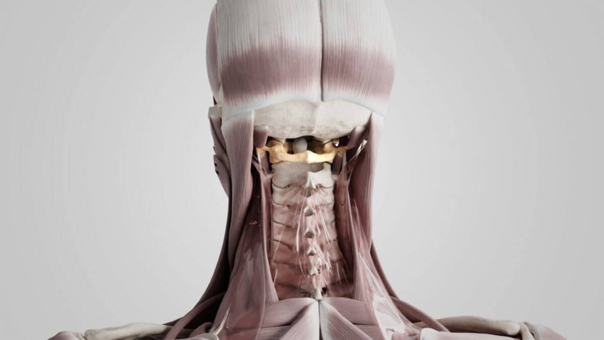
Corpus: Atlas
Synonym: C1, first cervical vertebra
1. Definition
The atlas is the first cervical vertebra (C1) and represents the part of the spinal column closest to the skull. Together with the second cervical vertebra, the axis (C2), it forms a functional unit and is also known as the "nodding vertebra," as it allows the head to flex anteriorly.
2. Anatomy
2.1. Basic structure
The structure of the atlas differs significantly from that of other vertebrae. It has a ring-like shape, having lost its vertebral body during development. Instead, there are lateral and ventral bony thickenings known as the lateral masses. Two nearly semicircular bone arches, the atlas arches, extend from these masses:
- Ventral: anterior arch (arcus anterior atlantis)
- Dorsal: posterior arch (arcus posterior atlantis)
The atlas does not have a pronounced spinous process; instead, there is an elevation on the dorsal side of the posterior arch called the posterior tubercle. Similarly, an anterior tubercle is found on the ventral side of the anterior arch.
Lateral to the lateral masses are the transverse processes, which are developmental remnants of the costal processes and contain the transverse foramina typical of cervical vertebrae.
2.2. Joint surfaces
The articular surfaces that constitute the atlantooccipital joint with the occipital bone are located on the upper side of the lateral masses, known as the superior articular surfaces.
The articular surfaces to the axis form the atlantoaxial joint and are located on the lower side and the anterior inner side of the atlas. The inferior articular surfaces point downwards. The dens axis articulates with an oval cartilaginous surface in a depression on the back of the anterior arch of the atlas, known as the fovea dentis.
2.3. Foramina
The transverse foramen of the atlas is traversed by the vertebral artery, which enters the skull through the foramen magnum. The vertebral foramen of the atlas is divided into two sections by the transverse ligament of the atlas. The dens axis lies ventral to the ligament, while the spinal cord is dorsal.
3. Clinic
3.1. Developmental disorders
During embryonic development, disorders can occur in the formation of the atlas. This may result in partial fusion of the sclerotomes of the upper four somites, leading to an adhesion of the occipital bone to the atlas. This developmental disorder, known as atlas assimilation, can be complete or incomplete.
3.2. Misalignments and fractures
Misalignments can occur in the atlas, just as in other vertebrae. Given that the spinal cord runs directly through the atlas, misalignments can cause varying degrees of central nervous system disorders and affect the spine's alignment. This can also disrupt blood circulation and cerebrospinal fluid flow, leading to further complications.
A serious injury associated with the atlas is a neck fracture, particularly a fracture of the dens axis. This injury, though rarely unnoticed, poses significant danger due to the potential for the dens axis to injure or destroy the medulla oblongata if the transverse ligament of the atlas and the ligament of apex dentis are torn. Damage to the Medulla oblongata where parts of the respiratory center are located, can result in death within seconds. This injury is often seen in cases of hanging.
Another specific injury is the Jefferson fracture, a type of atlas fracture where the ring of the atlas is completely ruptured due to strong longitudinal forces.


