Corpus: Spinal column


Synonyms: backbone, spine
1. Definition
The spinal column is a vital part of the skeleton. It provides flexible support for the body and bears the weight of the head, neck, torso, and upper limbs. It also houses the spinal cord, a crucial part of the central nervous system. The spinal column is a defining structural feature of vertebrates.
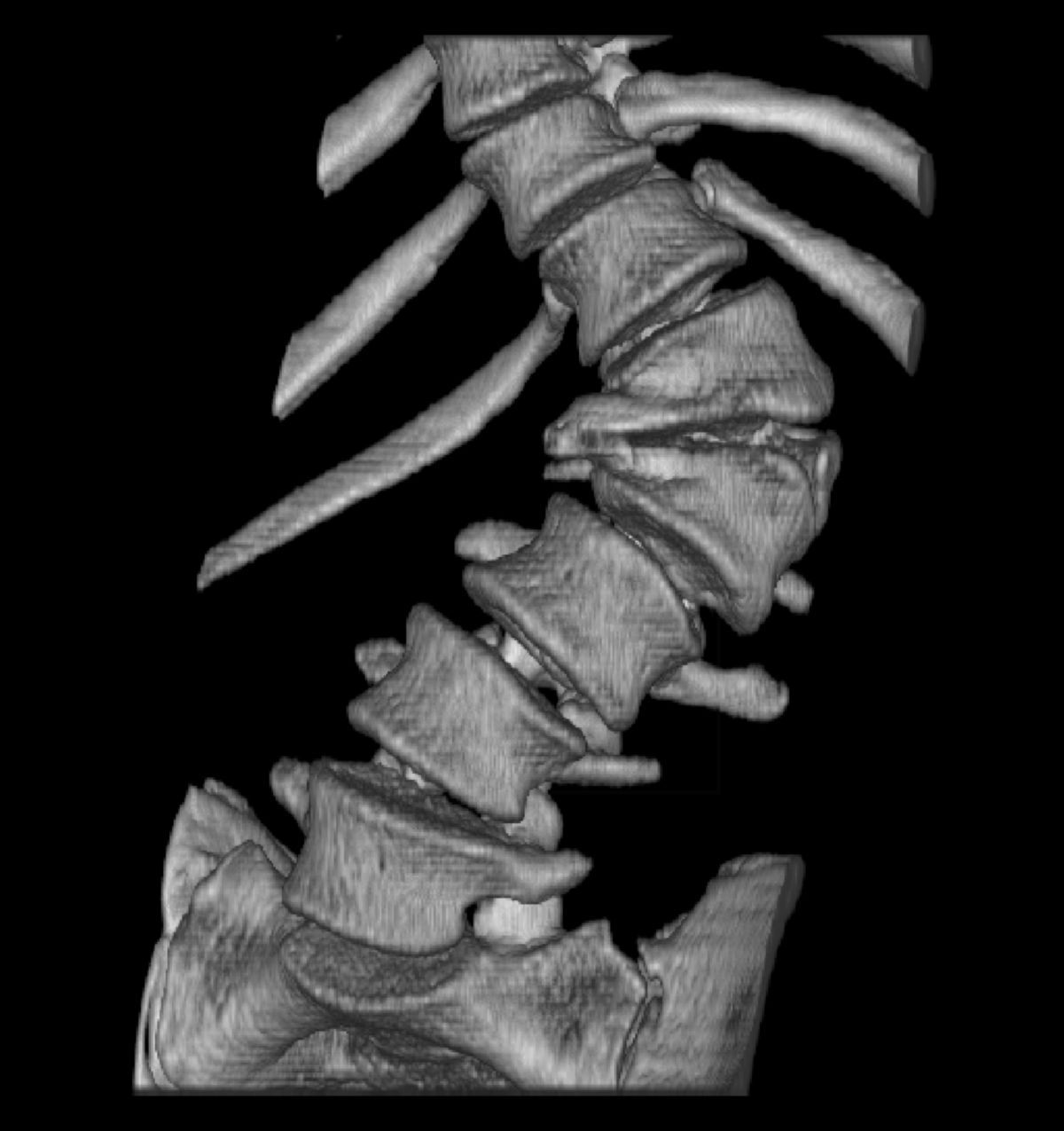
The individual segments of the spinal column are called vertebrae. The human spine consists of 24 vertebrae, which are connected by intervertebral discs and various ligaments.
2. Shape of the spinal column
When viewed from the side, the human spine has a double S-shape:
- Lordosis occurs in the cervical (neck) and lumbar (lower back) regions, where the spine curves forward (convex ventrally or concave dorsally).
- Kyphosis is found in the thoracic (mid-back) and sacral (pelvic) regions, where the spine curves backward (concave ventrally or convex dorsally).
When viewed from the front or back, the spine appears almost straight. A sideways curvature of the spine is called scoliosis. The normal shape of the spine is maintained by ligaments connecting the vertebrae and by the active support of the back muscles.
The spine, acting like a flexible spring, allows humans to stand and walk upright. It also helps absorb shocks caused by movement, protecting the brain from impact.
3. Division
The spinal column is divided into five sections:
- Cervical spine: 7 cervical vertebrae
- Thoracic spine: 12 thoracic vertebrae
- Lumbar spine: 5 lumbar vertebrae
- Sacrum: Formed by the fusion of 5 sacral vertebrae
- Coccyx: Consists of 4 to 5 coccygeal vertebrae that form the tailbone
4. Basic structure of the vertebrae
Each vertebra has three main parts: the vertebral body, vertebral arch, and various processes.
4.1. Vertebral body
The vertebral body is made up of spongy bone surrounded by a dense outer layer of compact bone. The upper and lower surfaces are called the superior and inferior plates, and they are connected by intervertebral discs.
4.2. Vertebral arch
This part extends from the back of the vertebral body and forms a protective ring around the spinal cord, creating the vertebral foramen. Together, all these foramina form the spinal canal. Notches in the vertebral arch create openings (intervertebral foramina) through which spinal nerves exit.
4.3. Vertebral processes
- Transverse processes: Extend laterally from the vertebral arch.
- Spinous process: Extends dorsally (toward the back).
- Superior and inferior articular processes: These form intervertebral joints, allowing movement between vertebrae.
5. Features of the vertebral structure
5.1. 1st cervical vertebra (atlas)
The atlas is a ring-shaped vertebra that lacks a vertebral body and spinous process. It is made up of:
- anterior arch of the atlas with the fovea dentis on the inside and the tuberculum anterius atlantis is on the outside
- Posterior arch of the atlas with a spinous process rudiment
- Lateral masses between the anterior and posterior arch of the atlas
The articular surfaces of the atlas are
- 2 superior articular surfaces
- 2 inferior articular surfaces
- 1 Fovea dentis
The two superior articular surfaces form the atlantooccipital joint together with the occipital bone.
5.2. 2nd cervical vertebra (axis)
The 2nd cervical vertebra, also known as the axis, has the largest bone mass among the cervical vertebrae. Its main feature is the dens axis, a peg-like structure, that forms the atlantoaxial joint with the atlas. Its articular surfaces include:
- anterior articular surface on the dens
- posterior articular surface on the dens
- two superior articular surfaces on the vertebral arch
- two inferior articular surfaces on the vertebral arch
5.3. Chest vertebrae
The key features of the thoracic vertebrae are the articular surfaces, which articulate with the ribs. Their distinctive structures include:
- two superior and inferior articular processes
- two superior and inferior costal facets ans the vertebral body
- two transverse costal facets on the transverse process
5.4. Lumbar vertebrae
Lumbar vertebrae are characterized by their large size and a smaller sagittal (front-to-back) diameter compared to their transverse diameter. Unique features include:
- costal processes: rib rudiments that replace the transverse processes
- accessory processes: small bony projections representing the true transverse processes
- mammillary processes: found on the back of the upper articular processes
5.5. Os sacrum
The sacrum is formed by the fusion of five sacral vertebrae into a triangular-shaped bone. Its key regions include:
- Base of the sacrum: broad upper part of the bone
- Apex of the sacrum: pointed lower end that articulates with the coccyx.
- Pelvic surface: front side of the sacrum, characterized by transverse ridges and anterior sacral foramina
- Dorsal surface: back side of the sacrum with median sacral crest (corresponding to spinous processes), medial sacral crest (corresponding to articular processes), lateral sacral crest (corresponding to transverse processes) and posterior sacral foramina
- Lateral part: also called the alae of the sacrum, including the auricular surface
The sacral canal is a continuation of the spinal canal, which opens at the bottom through the sacral hiatus.
5.6. Os coccygis
The coccyx is the lowest part of the spine, consisting of 4–5 small coccygeal vertebrae. These vertebrae are connected by cartilage (synchondroses) and eventually fuse together (synostosis) in adulthood.
6. Intervertebral discs (Disci intervertebrales)
Intervertebral discs are fibrocartilaginous structures located between the vertebral bodies. They consist of a tough, fibrous outer layer made of collagen fibers, called the annulus fibrosus. The annulus fibrosus surrounds a soft, gelatinous core, the Nucleus pulposus. It acts as a cushion and absorbs shocks.
Together with the vertebral bodies, the intervertebral discs form the intervertebral symphysis.
7. Ligaments of the spinal column
- Between the vertebral bodies
- Posterior longitudinal ligament: Runs along the back surface of the vertebral bodies.
- Anterior longitudinal ligament: Covers the front surface of the vertebral bodies.
- Between the vertebral arches:
- Between the transverse and spinous processes:
- Intertransverse ligaments: connect adjacent transverse processes
- Interspinous ligaments: connect the spinous processes
- Supraspinous ligament: links the tips of the spinous processes
- Ligamentum nuchae: between the occipital bone and the supraspinous ligament
- Between sacrum and coccyx:
- Superficial posterior sacrococcygeal ligament
- Deep posterior sacrococcygeal ligament: continuation of the posterior longitudinal ligament
- Anterior sacrococcygeal ligament: continuation of the anterior longitudinal ligament
- Lateral sacrococcygeal ligaments
8. Blood supply
The venous drainage from the vertebral bodies and the surrounding ligamentous apparatus occurs via the external vertebral venous plexus and the internal vertebral venous plexus.
9. Clinic
Diseases of the spine fall under the speciality of orthopaedics. They can be caused by traumatic, degenerative or other causes.
- Spinal deformities
- Scoliosis
- Gibbus
- Wedge vertebrae
- Spinal trauma
- Vertebral fracture
- Cervical spine distortion (whiplash injury)
- Degenerative diseases of the spine
- Osteoporosis
- Intervertebral disc prolapse
- Spinal tumours
- Bone metastases of the spine
- Vertebral body haemangiomas