Corpus: Cervical spine


Synonym: cervical vertebrae
1. Definition
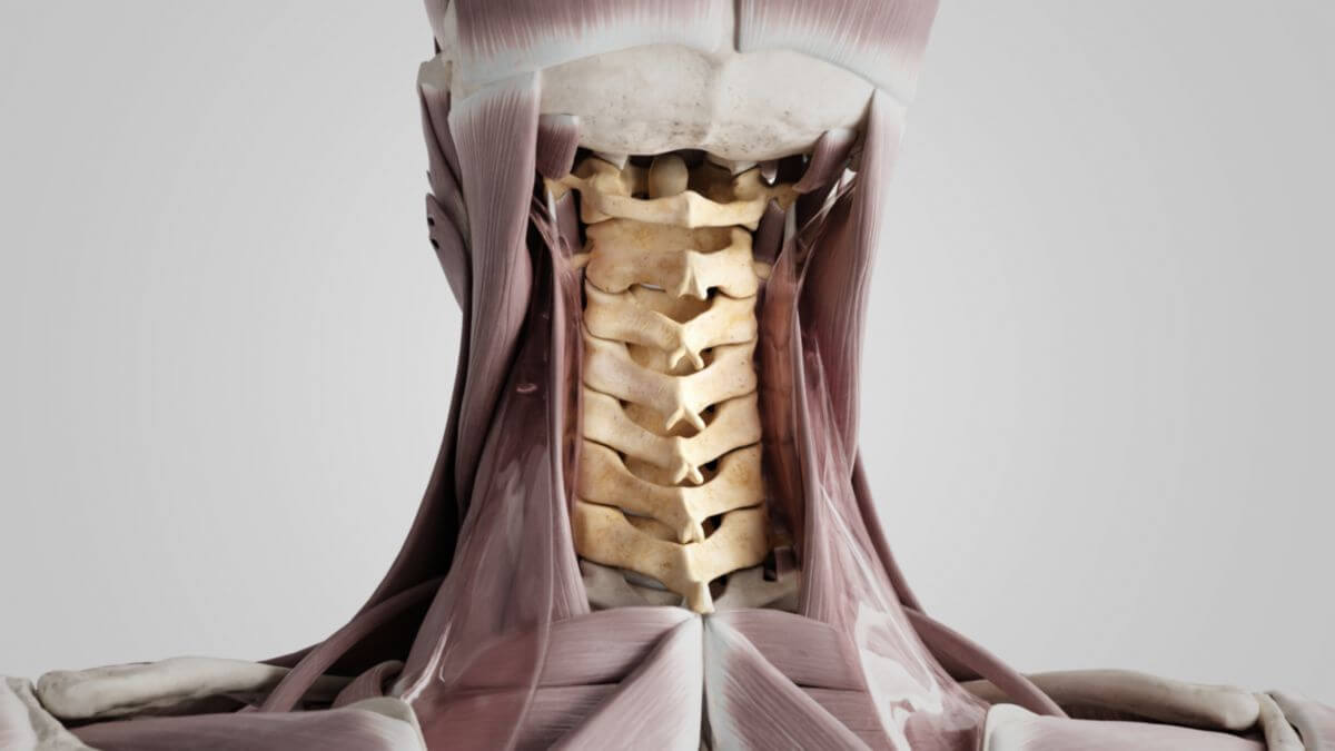
The cervical spine is the uppermost section of the spine located in the neck. It is the most mobile part of the spine and consists in humans of 7 cervical vertebrae (vertebrae cervicales). It continues caudally into the thoracic spine.
2. Overview
The cervical vertebrae are numbered from cranial (top) to caudal (bottom). Some vertebrae have specific names due to their unique shapes.
| Name | Acronym | Latin | Proper name |
|---|---|---|---|
| 1st cervical vertebra | C1, HWK 1 | Vertebra cervicalis I | Atlas |
| 2nd cervical vertebra | C2, HWK 2 | Vertebra cervicalis II | Axis |
| 3rd cervical vertebra | C3, HWK 3 | Vertebra cervicalis III | |
| 4th cervical vertebra | C4, HWK 4 | Vertebra cervicalis IV | |
| 5th cervical vertebra | C5, HWK 5 | Vertebra cervicalis V | |
| 6th cervical vertebra | C6, Cervical vertebra 6 | Vertebra cervicalis VI | |
| 7th cervical vertebra | C7, HWK 7 | Vertebra cervicalis VII | Vertebra prominens |
3. Anatomy
The seven cervical vertebrae are aligned in a column. In an upright posture, they form a dorsally concave arch known as cervical lordosis.
The first cervical vertebra, called the atlas, and the second, called the axis, along with the occipital bone, constitute the upper and lower head joints (atlanto-occipital and atlanto-axial joints). This area, along with adjacent cranial structures, ligaments, and muscles, is clinically referred to as the craniocervical junction.
Eight spinal nerves emerge from the spinal cord in the cervical spine region on each side. The nerves from segments C1 to C4 form the cervical plexus, while those from segments C5 to C8 form the brachial plexus.
3.1. Basic structure
Most vertebrae share a common structure with the following elements:
- Vertebral body
- Vertebral arch
From here, the lateral and dorsal processes of the vertebra (processus vertebrae) extend:
- Lateral: Transverse processes (processus transversi)
- Dorsal: Spinous processes (processus spinosi)
The vertebral foramen, enclosed by the vertebral body and arch, forms the space for the spinal cord (medulla spinalis) along with its sheaths, vessels, and nerves. The vertebral canal is formed by the sequential alignment of individual vertebral foramina. The intervertebral foramen, open between two vertebrae, allows passage of spinal nerves.
3.2. Special features
Cervical vertebrae are characterized by:
- A large, triangular vertebral foramen
- Inclined, almost horizontal articular processes
- Foramina transversaria in the transverse processes for the passage of the vertebral artery, vertebral veins, and sympathetic nerve fibers (C1-C6)
- Transverse processes with anterior and posterior tuberosities, with the sulcus nervi spinalis between them (C2-C7)
- Bifid spinous processes (C2-C6)
The atlas (1st cervical vertebra) differs from other vertebrae, resembling a ring with anterior and posterior arches, creating a pivot joint with the axis (2nd cervical vertebra) capable of 30° rotations on both sides. Unlike other vertebrae, the atlas lacks a spinous process, having only a posterior tubercle instead.
The vertebra prominens (7th cervical vertebra) marks the transition to the thoracic spine with a prominent spinous process that is easily palpable in the neck. Its transverse foramina are smaller compared to C1-C6 vertebrae.
Minor differences exist between the 3rd and 6th cervical vertebrae. By age ten, uncovertebral joints can develop between the 3rd and 7th cervical vertebrae.
3.3. Varieties
Variations in the cervical spine include:
- Accessory ossicle of the anterior arch of the atlas
- Sesamoid bone of the nuchal ligament
- Ossiculum terminale persistens
- Os odontoideum
4. Biomechanics
The cervical spine is the most mobile section of the spine, particularly in rotation, due to the position of articular surfaces in the intervertebral joints. It allows:
- Flexion and extension
- Lateral flexion (side bending)
- Rotation
These movements are controlled by the neck and back muscles.
5. Clinic
5.1. Diagnostics
Clinical and neurological examinations, along with imaging techniques, are essential for assessing the cervical spine. These include conventional cervical spine imaging (2 or 4 planes), CT scans, MRI, bone scintigraphy, discography, and myelography.
5.2. Diseases
The most important diseases of the cervical spine are based on trauma, degenerative changes or functional overload. These include, among others:
- Whiplash
- Cervical spine syndrome
- Torticollis
- Cervical radiculopathy
- Cervical disc herniation
- Ossification of the posterior longitudinal ligament
- Cervical spondylogenic myelopathy
- Atlantooccipital dislocation (AOD)
- Atlantoaxial subluxation (AASL)
- Syringomyelia
5.3. Malformations
Malformations of the cervical spine with fusion of vertebral bodies occur in Klippel-Feil syndrome.
6. Veterinary medicine
All pets also have seven cervical vertebrae.