Corpus: Thoracic spine


Synonym: thoracic vertebrae
1. Definition
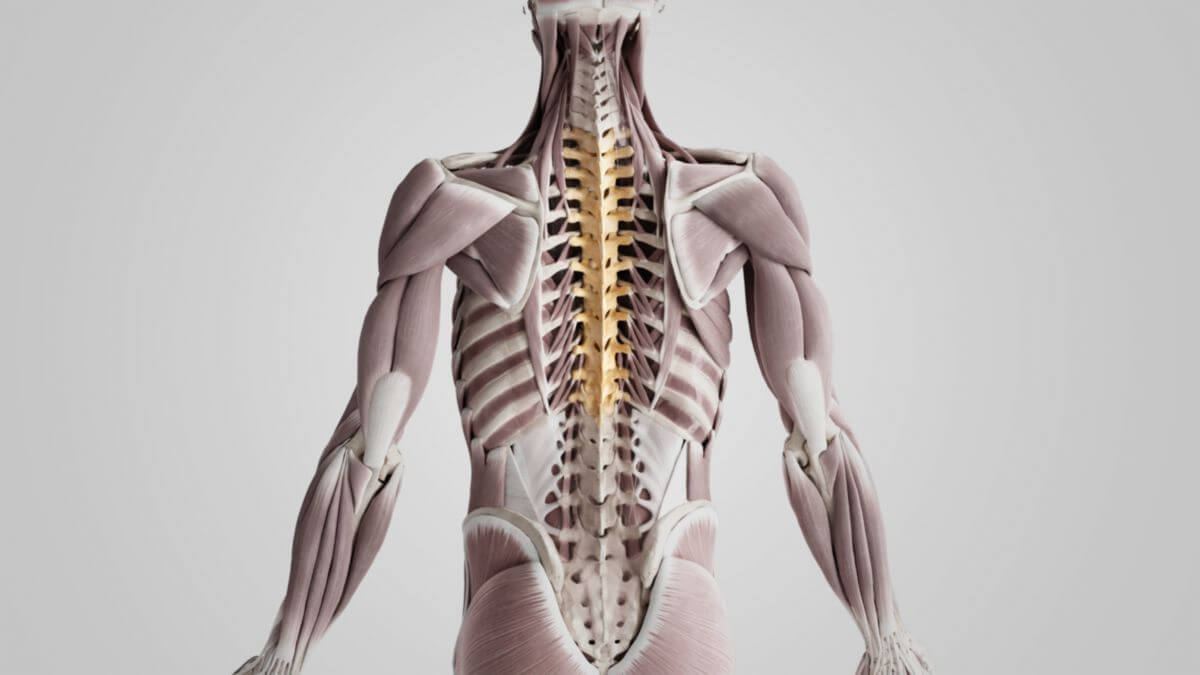
The thoracic spine is a segment of the human spine consisting of 12 individual bones known as the thoracic vertebrae. The thoracic spine connects to the ribs, forming the central structural unit of the thorax, and is situated between the cervical spine and the lumbar spine.
2. Overview
The thoracic vertebrae are numbered sequentially from cranial to caudal.
| Name | Acronyms | Latin |
|---|---|---|
| 1st thoracic vertebra | T1, Th1 | Vertebra thoracica I |
| 2nd thoracic vertebra | T2, Th2 | Vertebra thoracica II |
| 3rd thoracic vertebra | T3, Th3 | Vertebra thoracica III |
| 4th thoracic vertebra | T4, Th4 | Vertebra thoracica IV |
| 5th thoracic vertebra | T5, Th5 | Vertebra thoracica V |
| 6th thoracic vertebra | T6, Th6 | Vertebra thoracica VI |
| 7th thoracic vertebra | T7, Th7 | Vertebra thoracica VII |
| 8th thoracic vertebra | T8, Th8 | Vertebra thoracica VIII |
| 9th thoracic vertebra | T9, Th9 | Vertebra thoracica IX |
| 10th thoracic vertebra | T10, Th10 | Vertebra thoracica X |
| 11th thoracic vertebra | T11, Th11 | Vertebra thoracica XI |
| 12th thoracic vertebra | T12, Th12 | Vertebra thoracica XII |
3. Anatomy
The thoracic spine is composed of 12 vertebrae that are stacked in a rod-like structure. When in an upright posture, these vertebrae form a dorsally convex arch, creating a physiological curvature known as thoracic kyphosis.
Twelve pairs of spinal nerves originate from the thoracic spine. The spinal nerves from the Th1 segment contribute to the brachial plexus, while the nerves from the Th12 segment are involved in the lumbar plexus.
3.1. Basic shape
Each thoracic vertebra has a common structural design, with a vertebral body (corpus vertebrae) and a vertebral arch (arcus vertebrae). The lateral and dorsal processes of each vertebra include transverse processes (processus transversi) on the sides and spinous processes (processus spinosi) on the back. The spinous processes in the thoracic region are particularly easy to palpate.
The vertebral foramen, enclosed by the vertebral body and arch, forms the space for the spinal cord (medulla spinalis), along with its protective sheaths, blood vessels, and nerves. The collective arrangement of vertebral foramina creates the vertebral canal (canalis vertebralis), through which the spinal cord runs. The intervertebral foramina, located between adjacent vertebrae, allow the passage of spinal nerves.
3.2. Special features
Thoracic vertebrae differ in size and shape compared to cervical and lumbar vertebrae. The vertebral foramen in the thoracic region is nearly round, with the smallest diameter occurring between the 5th and 6th thoracic vertebrae. The spinal cord within the thoracic spine is the thinnest part of the entire spinal cord.
The pedicles (pediculi arcus vertebrae) of the thoracic vertebrae are also distinct, with variations in their cortical bone thickness according to the stress they bear. This characteristic is utilized in stabilizing surgical procedures, where long metal screws are inserted.
Scoliosis, a condition where the spine curves abnormally, is often most pronounced in the thoracic spine.
3.3. Joints
The thoracic vertebrae are involved in two main types of joints: facet joints (articulationes zygapophysiales) and costovertebral joints (articulationes costoverbrales).
3.3.1. Facet joints
The thoracic vertebrae have superior and inferior articular surfaces, which enable the connection to the neighboring vertebrae. These articular surfaces are located on the flat portion of the vertebral arch. Each thoracic vertebra has two articular processes extending cranially and two extending caudally. These processes form vertebral arch joints, also known as facet joints, which connect each vertebra with its cranial (upper) and caudal (lower) neighbors.
3.3.2. Costovertebral Joints
The thoracic vertebrae, in conjunction with the rib heads (capita costae), form the costovertebral joints. These joints are divided into two types based on their location:
- Joints of the rib heads: These are the joints between the rib head and the vertebral body.
- Costotransverse joints: These are the joints between the neck of the rib and the transverse processes of the thoracic vertebrae.
The articular surfaces for the rib heads are formed by the superior and inferior cosstal facets, which are articular depressions found on two adjacent thoracic vertebrae. Exceptions to this are the 1st, 11th, and 12th ribs, which have only one articular connection with the body of the respective vertebra.
The articular surfaces for the rib necks are located on the transverse processes of the 1st to 10th thoracic vertebrae, where they articulate with the costal tubercles of the ribs. From the 2nd to the 5th thoracic vertebrae, these articular surfaces are concave, while they are flat on the 1st, 6th, and 10th thoracic vertebrae. The articular surfaces for the rib necks are absent on the 11th and 12th thoracic vertebrae. Additionally, the 12th thoracic vertebra has a transverse process with unique features, including additional structures known as the mammillary process and accessory process, which are also found in lumbar vertebrae.
3.4. Ligaments
The thoracic spine is stabilized by a network of ligaments that support both the vertebral arch joints and the costovertebral joints. The ligaments specific to the thoracic spine include:
- Radiate ligament of the rib head: This ligament extends from the lateral aspect of the vertebral body to the head of the rib. It is most prominent in the 2nd to 10th ribs.
- Costotransverse ligaments:
- Lateral part: This portion extends from the transverse process of a thoracic vertebra to the tubercle of the same rib.
- Superior part: This portion extends from the transverse process to the upper edge of the costal tubercle of the rib below.
- Intra-articular ligament of the rib head: This ligament lies within the joint between the rib head and the vertebral body, providing internal stabilization for the articulation.
4. Range of motion
4.1. Flexion and extension
Flexion (forward bending) and extension (backward bending) of the trunk in the sagittal plane are primarily facilitated by the thoracic spine, although the cervical and lumbar regions contribute to the total range of motion. During forward bending, the curvature of the thoracic spine increases, enhancing the natural kyphosis. In contrast, backward bending flattens the thoracic spine. The range of motion for flexion is approximately 45° forward, while for extension, it is about 26° backward.
4.2. Lateral flexion
Lateral flexion, or sideways bending of the spine, involves the thoracic vertebrae and can occur independently in the cervical and lumbar regions or as part of a coordinated motion involving the thoracic spine to create a full arch. The range of lateral flexion in the thoracic spine is between 25° and 33°.
4.3. Rotation
Rotation around the spine’s vertical axis is most prominent in the cervical spine. However, the thoracic spine contributes significantly to trunk rotation, allowing for approximately 33° of rotational movement.
5. Clinic
5.1. Diagnostics
Clinical evaluation of the thoracic spine is essential in diagnosing spinal abnormalities. This includes taking a detailed medical history, performing a visual inspection, and palpating the spinous processes and surrounding musculature to detect any irregularities or tenderness. X-ray imaging, particularly in frontal and lateral views, is crucial for assessing the spine. Additional diagnostic tools such as CT and MRI provide more detailed images of the spine, while skeletal scintigraphy is used to detect bone metastases, which frequently affect the thoracic vertebrae.
5.1.1. Simple orthopaedic examination
A basic orthopedic assessment of the thoracic spine starts with inspection, focusing on the shape, alignment, and position of the spinous processes. Palpation of the spinous processes and the entire spine follows, assessing for abnormalities or tenderness. The range of motion is evaluated using specific tests:
- Flexion and Extension: Assessed with the Ott sign, Schober sign, and finger-to-floor distance.
- Lateral Flexion: Measured using the finger-to-knee distance.
- Rotation: Evaluated by having the patient sit on a swivel chair and rotate their torso to the left and right, allowing the examiner to estimate the rotational angles.
6. Veterinary medicine
The number of thoracic vertebrae, and consequently the number of ribs, varies widely among animal species and even within breeds. For example:
- Carnivores: Typically have 12 to 14 thoracic vertebrae.
- Pigs: Have 13 to 16 thoracic vertebrae.
- Sheep and Goats: Usually have 13 thoracic vertebrae.
- Cattle: Commonly have 13 thoracic vertebrae.
- Horses: Have 18 thoracic vertebrae.
These variations influence the thoracic structure and function across different species.