Corpus: Brachial plexus


1. Definition
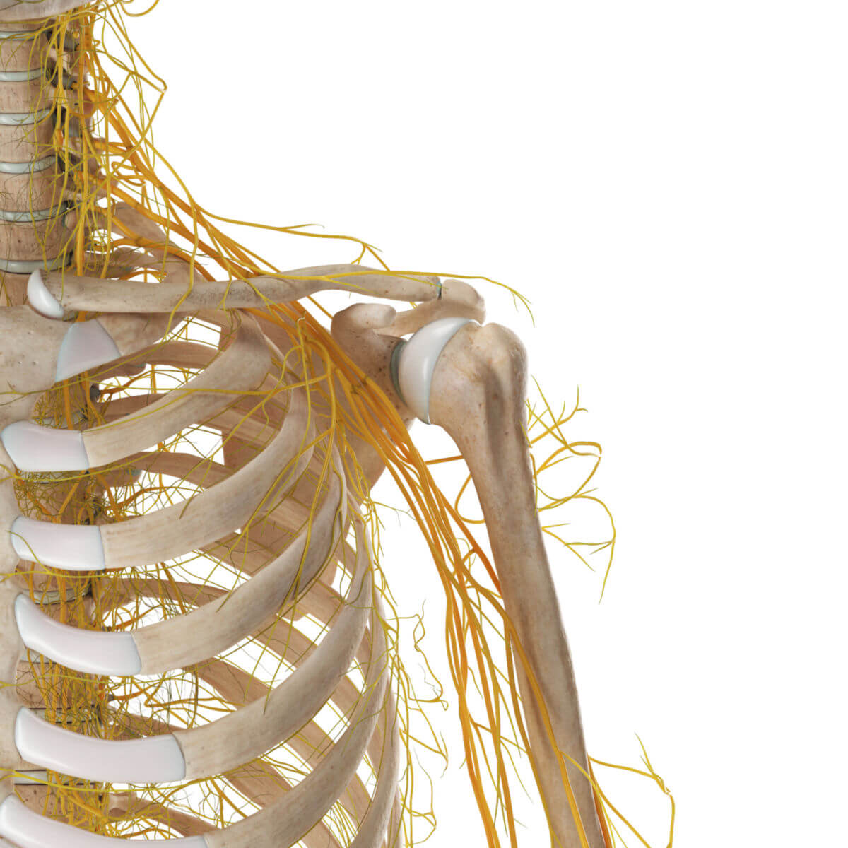
The brachial plexus is a network of nerves in the peripheral nervous system (PNS), formed by the anterior branches (rami) of the spinal nerves C5-C8 and T1. It gives rise to nerves that innervate the arm, shoulder, and chest.
2. Function
The brachial plexus provides motor innervation to the shoulder and chest muscles, as well as motor and sensory innervation to the arm and hand.
3. Embryology
The complex structure of the brachial plexus results from the outgrowth of the limb buds at the end of the 4th embryonic week. During this process, the myotomes, which were originally arranged segmentally, are displaced. The associated nerves (ventral rami of the spinal nerves C5-T1) follow the myotomes and intertwine to form the brachial plexus. The middle areas (e.g., C7) are pushed furthest distally during limb growth.
4. Structural organisation
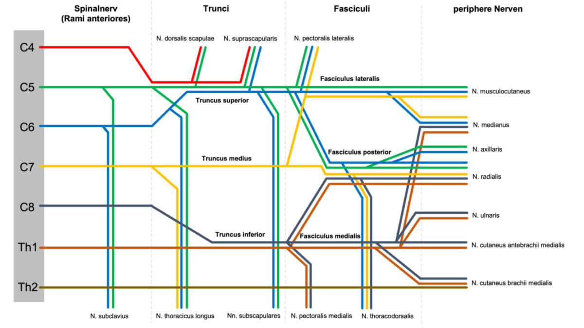
From a structural point of view, the brachial plexus can be roughly divided into three sections: the trunks (trunci), the cords (fasciculi), and the nerves (nervi).
4.1. Trunci
The trunci, or primary trunks, are the main trunks of the brachial plexus that directly follow the anterior branches of the spinal nerves and run between the scalene muscles through the vertical scalene gap. They are classified based on their topographical arrangement:
- Superior trunk: Arises from the union of nerve fibers from C5 and C6
- Middle trunk: Arises directly from the nerve fibers from C7
- Inferior trunk: Arises from the fusion of nerve fibers from C8 and T1
As an anatomical variation, smaller fiber portions of the fourth cervical segment (C4) and the second thoracic segment (T2) can sometimes be involved in the formation of the brachial plexus.
After passing through the scalene gap, the brachial plexus is covered by a coarse connective tissue sheath that accompanies it into the axilla. This sheath also contains the axillary artery and vein in addition to the nerves.
4.2. Fasciculi
Further distally, each trunk gives off an anterior and a posterior branch (anterior and posterior division), which in turn form the cordes or secondary trunks. These arrange themselves around the axillary artery, so the nomenclature of the fascicles refers to their position relative to the artery. The distinctions are:
- Lateral fasciculus: Formed by the union of the anterior divisions of the superior and middle trunks.
- Medial fasciculus: Arises from the anterior division of the inferior trunk.
- Posterior fasciculus: Formed from the posterior divisions of all three trunks.
4.3. End branches
The individual fasciculi give rise to the various nerves, formed by the fusion of fiber parts from different fascicles. They are briefly outlined here:
- Musculocutaneous nerve: Arises from the lateral fasciculus and supplies the motor flexors of the upper arm and a sensory area of the forearm.
- Median nerve: Formed by the fusion of fibers from the lateral and medial fasciculi; it innervates various flexors in the forearm, some muscles of the hand, and provides sensory innervation to the palm (excluding the little finger) and parts of fingers I-IV.
- Ulnar nerve: Arises from the medial fasciculus and innervates some flexors in the forearm, large parts of the hand muscles, and provides sensory innervation to the ulnar parts of the hand, the hypothenar eminence, and fingers IV (partially) and V.
- Medial antebrachial cutaneous nerve and medial brachial cutaneous nerve: These nerves originate from the medial fasciculus and provide sensory innervation to the medial side of the upper arm and forearm.
- Axillary nerve: Originates from the posterior fasciculus and innervates motor parts of the shoulder girdle muscles and provides sensory innervation to the skin of the shoulder.
- Radial nerve: Also originates from the posterior fasciculus and innervates all extensors of the arm, parts of the hand muscles, and provides sensory innervation to the dorsal side of the arm.
4.4. Other nerves
The following nerves arise directly from the spinal cord segments or from the trunks:
5. Topographical subdivision
The brachial plexus can be divided into two sections based on topographical aspects:
- Pars supraclavicularis: Located above the clavicle
- Pars infraclavicularis: Located below the clavicle
5.1. Pars supraclavicularis
The three trunks (superior, middle, and inferior) and the following nerves are found in the supraclavicular part:
- Dorsal scapular nerve (C4, C5)
- Subclavian nerve (C5, C6)
- Long thoracic nerve (C5-C7)
- Suprascapular nerve (C4-C6)
5.2. Pars infraclavicularis
The three fasciculi (lateral, medial, and posterior) of the brachial plexus are found in the infraclavicular part, lying in the deep layer of the infraclavicular fossa (Mohrenheim's fossa). They are named according to their relative position to the axillary artery. The fasciculi and their branches are detailed below:
- Lateral fasciculus
- Musculocutaneous nerve
- Median nerve (lateral root)
- Medial fasciculus
- Ulnar nerve
- Medial brachial cutaneous nerve
- Medial antebrachial cutaneous nerve
- Median nerve (medial root)
- Posterior fasciculus
- Radial nerve
- Axillary nerve
Shortly before the fascicles, the short infraclavicular branches supply the muscles of the shoulder:
- Thoracodorsal nerve (C6-C8)
- Subscapular nerves (C5, C6)
- Medial pectoral nerves (C8, T1) and lateral pectoral nerves (C5-C7)
Cave: There are numerous anatomical variations of the nerve branches. The above description represents one possible configuration.
6. Situs
7. Clinic
7.1. Diseases
Diseases of the nerve plexuses are referred to as plexopathies, or plexus lesions, if caused by trauma. Following a partial or complete rupture of the nerve roots of the brachial plexus due to violence (e.g., traction) or birth trauma, a condition known as brachial plexus palsy can occur. It is characterized by sensory and motor deficits in the arm. There are two types:
- Upper plexus paralysis (Erb-Duchenne type): Affects segments C5 and C6.
- Lower plexus paralysis (Klumpke type): Affects segments C7, C8, and T1.
Narrowing of the passage of the brachial plexus through the posterior scalene gap in the neck can lead to scalene syndrome.
Neuralgic amyotrophy is an inflammation of the brachial plexus triggered by circulating immune complexes.
Rarely, tumors such as schwannomas, neurofibromas, and malignant peripheral nerve sheath tumors (MPNST) can originate from the brachial plexus.
7.2. Diagnostics
The brachial plexus can be examined indirectly through neurological functional testing of the nerves it forms, and directly via neurophysiological diagnostics and neuroradiological imaging. MR neurography with high-resolution MR systems or 3D ultrasound are used for visualization.
7.3. Anesthesia
The brachial plexus can be selectively anesthetized using local anesthetics during surgical procedures on the arm or for pain management. This form of regional anesthesia is called plexus anesthesia or plexus blockade. There are different types of blocks:
- Interscalene plexus blockade
- Axillary plexus block
- Supraclavicular plexus blockade
- Infraclavicular plexus blockade
8. Memorising aids
To help remember the fascicles from which the individual nerves originate, you can use the following mnemonics:
| Mnemonic | Nerve | Fascicle |
|---|---|---|
| Marylin | Musculocutaneous nerve | Lateral fasciculus |
| Monroe | Median nerve | Lateral fasciculus and medial fasciculus |
| and | Ulnar nerve | Medial fasciculus |
| King | Nervus cutaneus brachii medialis | Medial fasciculus |
| Cong | Nervus cutaneus antebrachii medialis | Medial fasciculus |
| rescue the | Radial nerve | Posterior fasciculus |
| Anatomy | Axillary nerve | Posterior fasciculus |