Corpus: Median nerve


from Latin: medianus - in the centre
Definition
The median nerve is a mixed motor and sensory nerve of the arm that arises from the brachial plexus.
Course
The median nerve originates from the lateral cord and the medial cord of the brachial plexus. It contains fibers from segments C6 to Th1. Its two original branches wrap around the axillary artery (so-called "medianus loop") and run laterally of the brachial artery to the distal side. Approximately at the level of the insertion point of the coracobrachialis muscle, it crosses over or under the brachial artery in the medial bicipital sulcus and then lies medial to the vessel.
Via the medial side of the crook of the elbow, the median nerve continues under the bicipital aponeurosis to the forearm. There it is initially found between the two heads of the pronator teres muscle (humeral and ulnar head). This point of passage is also known as the median nerve tunnel.
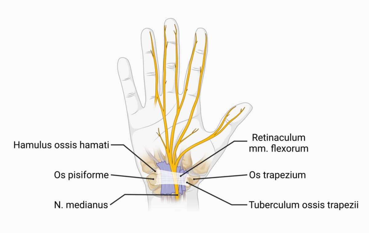
Further along, the nerve crosses the ulnar artery, from which it is separated by the deep head of the pronator teres muscle. Between the flexor digitorum superficialis and profundus muscles, it descends further to the wrist. 4-5 cm before the transverse carpal ligament, the nerve rises from the depths of the forearm and then lies between the tendons of the flexor digitorum superficialis muscle and the flexor carpi radialis muscle - partially overlaid by the tendon of the palmaris longus muscle. At this point it is only covered by fascia and skin. Under the transverse carpal ligament, it finally passes through the carpal canal into the palm.
Branches on the Forearm
With the exception of a branch to the pronator teres muscle, which sometimes branches off proximal to the elbow, the median nerve does not give off any branches on the upper arm. It has the following branches on the forearm:
- muscular branches
- palmar branch of median nerve
Branches on the Hand
In the palm of the hand, the median nerve is divided into a lateral and a medial branch, which send the following nerves to the fingers:
Anastomoses
The connection between the median nerve and the ulnar nerve on the forearm is known as Martin-Gruber anastomosis. It is a common anatomical variant.
Function
Motor Innervation
The median nerve innervates almost all flexors of the forearm muscles. The only exceptions are the flexor carpi ulnaris muscle and the ulnar part of the flexor digitorum profundus muscle (both innervated by the ulnar nerve). It also supplies all the pronators. The nerve is therefore primarily responsible for flexion of the fingers and hand as well as pronation. It also supplies parts of the muscles of the ball of the thumb (thenar muscles) and two of the short muscles of the metacarpus.
Forearm
- flexor carpi radialis muscle
- flexor digitorum profundus muscle, radial part
- flexor digitorum superficialis muscle
- palmaris longus muscle
- pronator teres muscle
Hand
- flexor pollicis brevis muscle, superficial part
- lumbricales muscles (first and second)
Sensitive Innervation
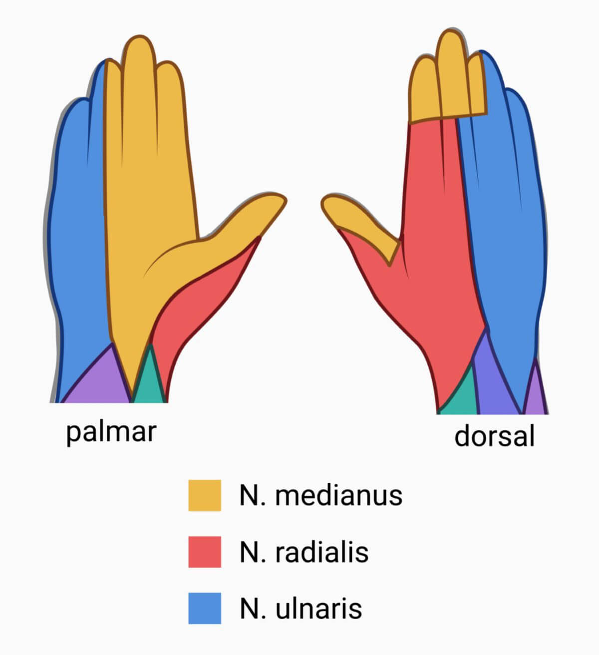
On the palmar surface of the hand, the median nerve sensitively supplies the skin over the ball of the thumb and the radial part of the palm. On this side, it also supplies the skin of the thumb, index and middle finger, as well as the radial half of the ring finger. On the dorsal side of the hand, it supplies the distal ends of fingers I-III from the proximal interphalangeal joint, as well as the radial half of the ring finger end phalanx.
Clinic
Paralysis of the median nerve is known as median nerve palsy. With this neurological disorder, it is no longer possible to close the fist. When attempting to close the fist, the thumb, index, and middle fingers can no longer be fully flexed, which is clinically manifested as a so-called preacher's hand. In addition, there is a loss of sensitivity in the supply areas described above. In the case of a proximal median nerve lesion, pronation weakness also occurs.
A common disease of the median nerve is carpal tunnel syndrome, with paraesthesia around the hand and - if it persists for a long time - atrophy of the thenar muscles.