Corpus: Pronator teres muscle

from latin: teres - round
1. Definition
2. Anatomy
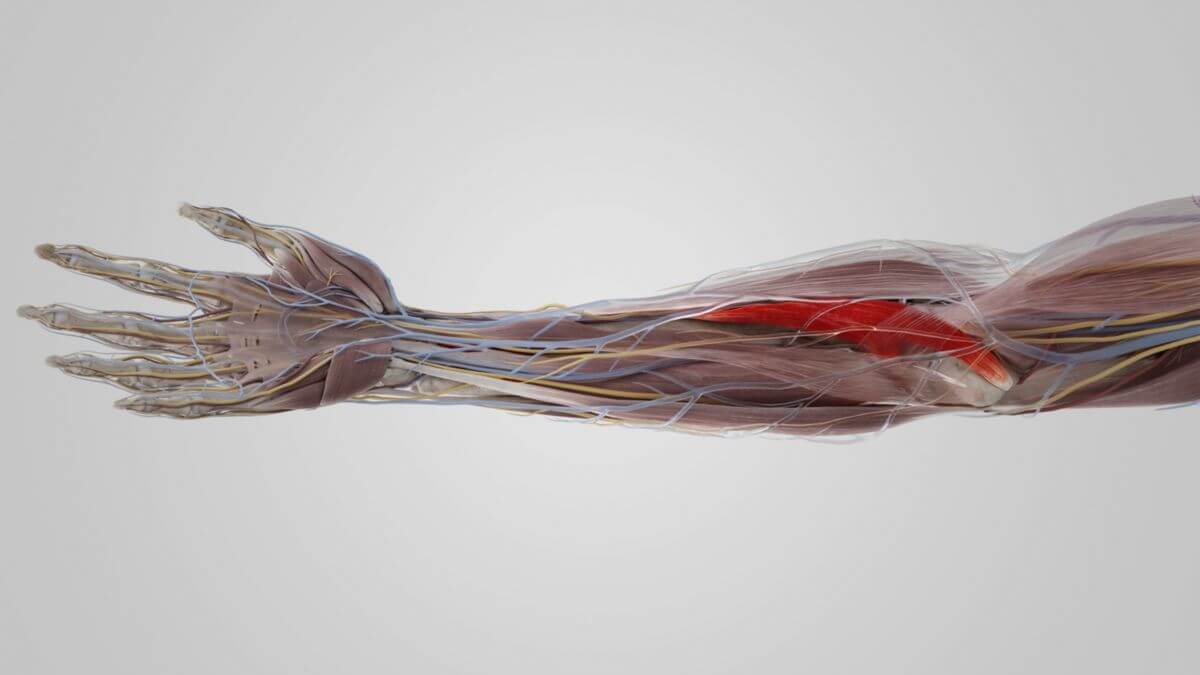
The humeral head of the pronator teres muscle originates from the medial epicondyle of the humerus and shares a common origin tendon with other forearm flexors. Additional origin sites include the forearm fascia (antebrachial fascia) and the muscle septum, which separates it from the flexor carpi radialis muscle.
The ulnar head is a relatively thin bundle of fibers originating from the coronoid process of the ulna and covers the ulnar artery at this location. The median nerve runs between the muscle heads in the pronator slit formed by both heads.
The pronator teres muscle runs obliquely across the forearm and has its tendinous insertion in the middle third of the radius, on the lateral face or the anterior margin of the bone.
The muscle forms the medial boundary of the cubital fossa.
3. Innervation
Like the other forearm flexors, the pronator teres muscle is innervated by the median nerve (segments: C6 and C7).
Since this nerve passes distally between the two heads of the muscle, the pronator teres muscle is the key muscle for the median nerve.
4. Function
Along with the pronator quadratus muscle, the pronator teres muscle is the most important muscle for pronation of the forearm. Additionally, it is responsible for flexion (bending) at the elbow joint along with the other forearm flexors.