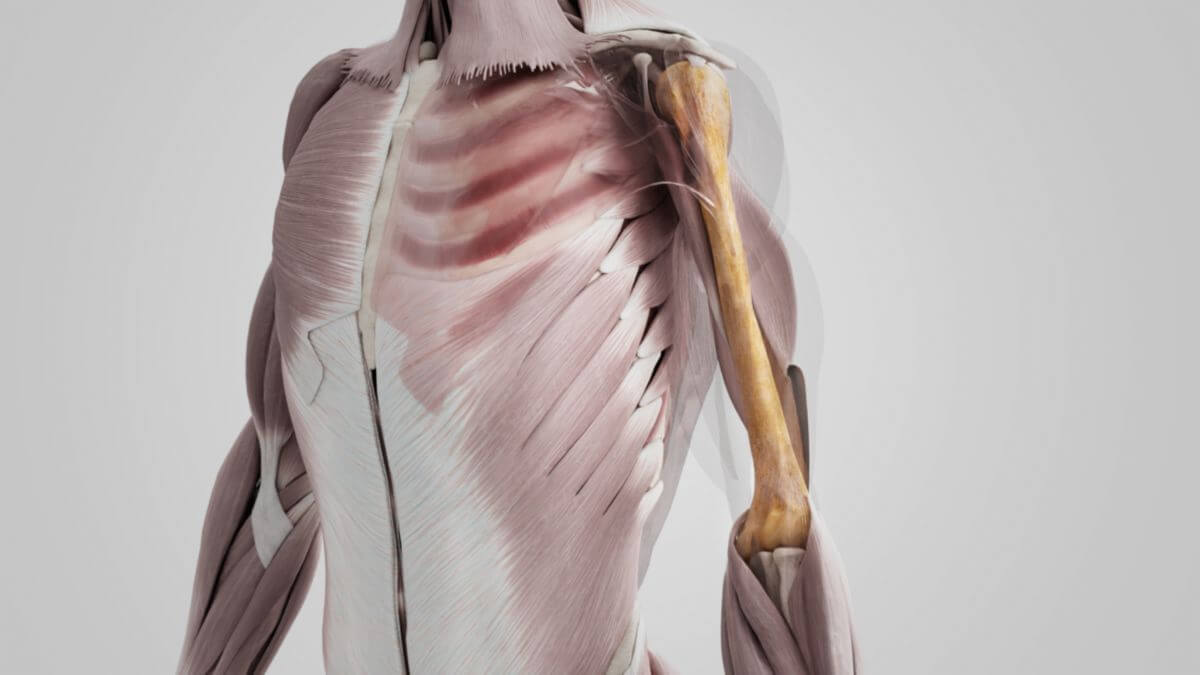
Corpus: Humerus


Synonym: upper arm bone
1. Definition
2. Extremitas proximalis
Four important anatomical structures can be distinguished at the end section facing the trunk, the extremitas proximalis: the head (caput humeri), the neck (collum humeri) and two tubercles, the tuberculum majus humeri and the tuberculum minus humeri.
2.1. Caput humeri
The almost hemispherical head of the humerus is directed cranially and medially as well as slightly dorsally when the humerus is at rest. It is positioned in relation to the shaft axis at a collodiaphyseal angle of 120 to 140° and a retrotorsional angle of 25 - 40°. Together with the glenoid cavity of the scapula, the humeral head forms the shoulder joint.
2.2. Collum humeri
Immediately below the humeral head is the neck of the humerus. It is slightly retracted in relation to the joint surface and is also known as the "collum anatomicum". It serves as an attachment for the joint capsule of the shoulder joint. The "collum chirurgicum", a surgically significant "predetermined breaking point" of the humerus, which lies below the tuberosity in the transition to the corpus, is separated from it.
2.3. Tuberculum majus (humeri)
The greater tuberosity lies lateral to the humeral head. Its cranial surface is rounded and has three flat impressions that serve as attachments for the following muscles from top to bottom:
The lateral surface of the greater tuberosity is rough and convex and merges into the corpus humeri without a visible boundary.
2.4. Tuberculum minus (humeri)
The lesser tuberosity also serves as a muscle insertion for the subscapularis muscle. Although it is smaller than the greater tuberosity, it protrudes more anteriorly. It points medially, in the direction of pull of the tendon of the subscapularis muscle.
2.5. Intertubercular sulcus
Both tubercles end caudally in a bone ridge, the crista tuberculi majoris and the crista tuberculi minoris. A groove lined with fibrous cartilage, the intertubercular sulcus, runs between the tuberosities. It extends distally to approximately the beginning of the middle third of the bone and becomes increasingly less pronounced. The sulcus is closed by the transverse ligament of the humerus to form an osteofibrous canal. The long tendon of the caput longum of the biceps brachii muscle and a branch of the anterior circumflex humeral artery run through it.
3. Corpus humeri
The actual body or shaft of the humerus (corpus humeri) has various surfaces (facies) and edges (margins).
3.1. Surfaces
3.1.1. Facies anterior lateralis
The proximal part of the facies anterior lateralis is smooth and rounded. It is covered by the deltoid muscle. Approximately in the center of the surface there is a rough hump, the deltoid tuberosity, which marks the insertion of the muscle of the same name. Below it is the sulcus nervi radialis coming from the posterior facies, in which the radial nerve and the profunda brachii artery swivel from diagonally behind and above to the front. In the distal part, the facies anterior lateralis serves as the origin of the brachialis muscle.
3.1.2. Facies anterior medialis
The facies anterior medialis is less extensive than the facies anterior lateralis. Its upper part is narrow, forms the base of the intertubercular sulcus and serves as the attachment surface of the latissimus dorsi muscle. A rough area in the middle section forms the insertion of the coracobrachialis muscle. The distal part serves as the origin of the brachialis muscle.
3.1.3. Posterior facies
The posterior facies is almost completely covered by the lateral and medial head of the triceps brachii muscle. The surface of origin of both muscle heads is intersected by the radial sulcus.
3.2. Margins
3.2.1. Anterior margo
The anterior margo runs from the front of the greater tuberosity distally to the coronoid fossa and thus separates the medial anterior facies from the lateral anterior facies. In the upper part it is a prominent bony projection (crista tuberculi majoris), which serves as an attachment for the tendon of the pectoralis major muscle. In the center it takes up the anterior border of the deltoid tuberosity. Distally, it appears smooth and rounded and serves as the origin of the brachialis muscle.
3.2.2. Lateral margo
The lateral margo extends from the back of the greater tuberosity to the lateral epicondyle of the humerus, thereby separating the anterior lateral facet from the posterior facet. The upper half is rounded and difficult to delineate. It serves as the insertion for the teres minor muscle and, distally, as the origin for the lateral head of the triceps brachii muscle. In the center, the margo lateralis is crossed by the flat bony impression of the radial sulcus. Distally, it ends in a pronounced bony crest, the crista supracondylaris lateralis, which serves as the origin of the brachioradialis muscle.
3.2.3. Margo medialis
The margo medialis extends from the lesser tuberosity to the epicondylus medialis humeri. In the upper third, it appears as a prominent bony protrusion (crista tuberculi minoris). This is where the tendon of the teres major muscle attaches. In the middle it has a slight depression for the attachment tendon of the coracobrachialis muscle. The lower third rises to form a bony protrusion, the crista supracondylaris medialis, which becomes more pronounced as it approaches the epicondyle.
4. Extremitas distalis
The end section of the humerus facing away from the trunk, the extremitas distalis, is flattened in an anterior-posterior direction and forms the condyle of the humerus. It carries two articular surfaces for the elbow joint: laterally the humeral capitulum and medially the humeral trochlea. Laterally there are two bony projections, the medial humeral epicondyle and the lateral humeral epicondyle.
4.1. Capitulum humeri
The lateral part of the articular surface consists of a button-like rounded, hemispherical and clearly bulging cartilage surface, the humeral capitulum. It articulates with the concave articular surface of the radial head.
4.2. Trochlea
The medial part of the articular surface is called the trochlea humeri. It extends as a transverse cylinder from the front to the back of the humerus and thus forms an extended, waisted cartilage roll. The articular roller is concave in the mediolateral direction. The trochlea articulates flush with the corresponding, crescent-shaped articular surface of the olecranon of the proximal ulna. The lateral border separates the trochlea in the shape of a ridge from the adjacent cartilaginous surface, which is connected to the edge of the radial head. The medial border is more pronounced and marks the transition to the medial epicondyle.
4.3. Radial fossa
Just proximal to the capitulum humeri, you can see a depression in the bone, the radial fossa. It accommodates the front edge of the radial head when the forearm is brought into a strong flexion position.
4.4. Coronoid fossa
A small depression, the coronoid fossa, can be seen proximal to the trochlea in the anterior view. Analogous to the radial fossa, it accommodates the coronoid process of the ulna when the forearm is flexed.
4.5. Olecranon fossa
On the back of the humerus, proximal to the trochlea, there is another clearly defined triangular depression, the olecranon fossa. In the extended position of the forearm, it accommodates the olecranon of the ulna.
4.6. Lateral epicondyle
The lateral epicondyle is a small bony prominence with numerous tuberosities on the lateral side of the humerus. It is the common origin of the following muscles:
- supinator muscle
- extensor carpi radialis brevis muscle
- extensor carpi ulnaris muscle
- extensor digitorum muscle
- extensor digiti minimi muscle
- anconeus muscle
It also serves as an attachment surface for the fibers of the radial collateral ligament. Proximally, it continues into the lateral supracondylar crest.
4.7. Medial epicondyle
The medial epicondyle is more pronounced than the lateral one. Proximally, it continues into the medial supracondylar crest. It serves as an attachment surface for the ulnar collateral ligament. The medial epicondyle is the common origin of the superficial flexors of the forearm:
- pronator teres muscle
- flexor carpi radialis muscle
- flexor carpi ulnaris muscle
- palmaris longus muscle
- flexor digitorum superficialis muscle
The ulnar nerve runs in the ulnar nerve sulcus on the posterior side of the medial epicondyle.
5. Vascular supply
The arterial supply to the humerus is provided by several smaller vessels:
- anterior humeral circumflex artery artery from the axillary artery
- humeral nutrient arteries from the arteria profunda brachii
6. Development
The onset of diaphyseal ossification of the humerus begins in the 7th - 8th week of development. The bone nuclei in the epiphysis appear proximally between the 1st and 4th year of life. In the distal epiphysis, ossification begins in the humeral capitulum as early as 1 - 2, in the medial humeral epicondyle in the 5th-6th, and in the humeral trochlea and lateral humeral epicondyle in the 8th-13th year of life. The distal epiphyseal groove closes between the ages of 14 and 18, the proximal groove only between the ages of 20 and 25. Bone nucleation and epiphyseal joint closure take place somewhat earlier in females than in males. If the axis of the humeral head is projected from the middle of the greater tuberosity to the middle of the caput humeri onto the epicondylar axis of the elbow joint, the torsion angle is obtained. In newborns, it is about 60° (torsion of the humerus). The flattening of the thorax in the sagittal direction changes the position of the scapula. The humeral head must follow this change so that the torsion angle in adults is only around 20°. Length growth takes place mainly in the proximal epiphysis. The proximal epiphyseal groove is located in the area of the collum anatomicum until the age of five. After that, the epiphyses of the caput humeri and the greater tuberosity unite, so that the epiphyseal groove is displaced under the greater tuberosity ("secondary" epiphyseal groove). As a result, it loses the protection of the joint capsule and the rotator muscles. In the event of violent impact, it is now relatively easy for the epiphysis to shear off the shaft (epiphysolysis capitis femoris). In adolescence, the joint moves to the area of the tuberosity ("tertiary" joint) and is again better protected.
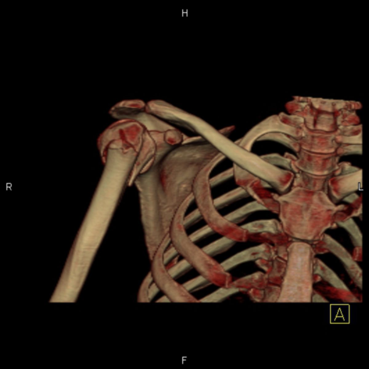
7. Clinic
Fractures of the humerus are relatively common due to the exposed position of the upper arm and account for approx. 4 – 5 % of all fractures. A distinction is made between:
- proximal humerus fracture (humeral head fracture)
- humeral shaft fracture
- distal humerus fracture
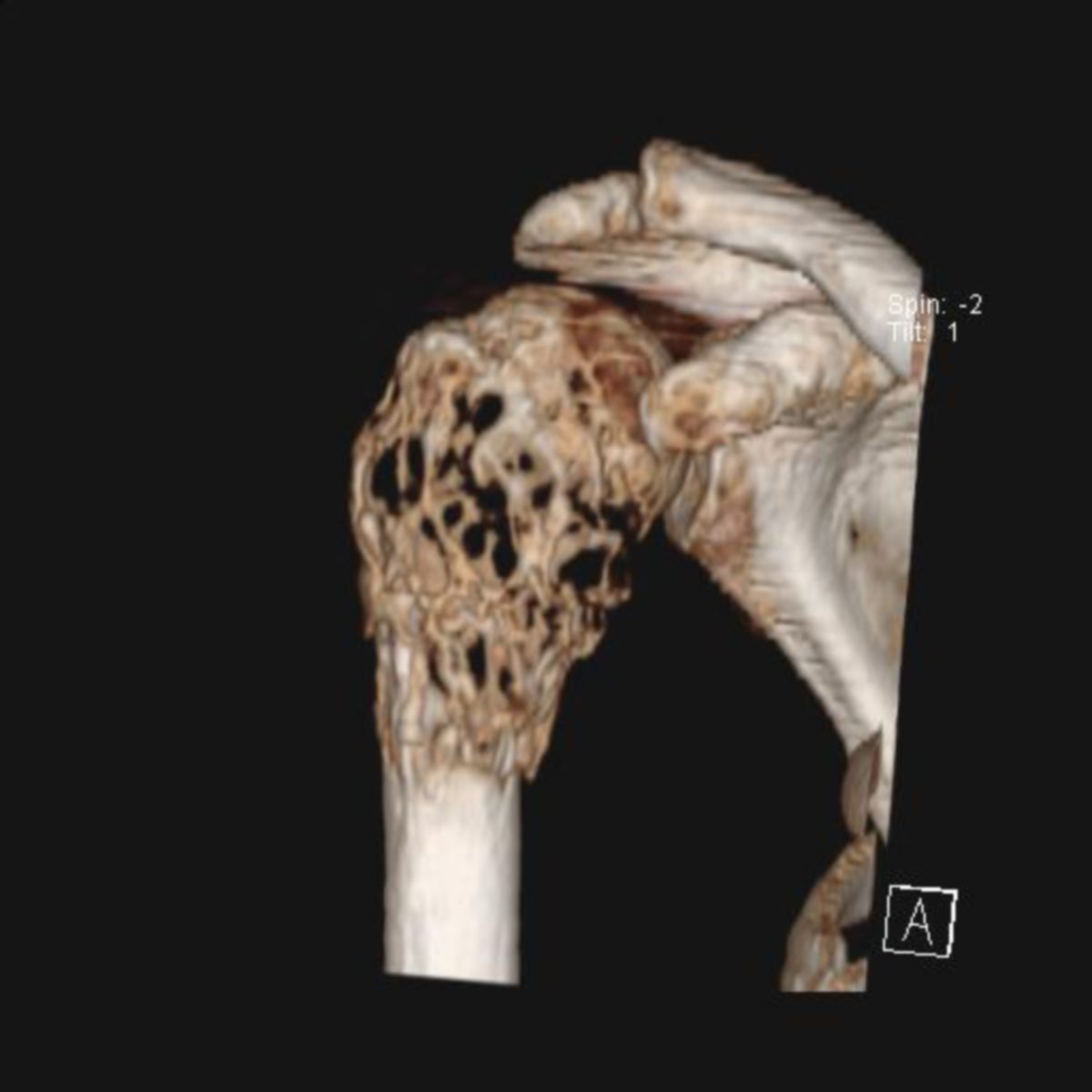
In rare cases, aseptic bone necrosis of the humerus, known as humeral head necrosis, can occur. Above the medial epicondyle, a hook-shaped projection, the supraepicondylar process, is found in around one percent of people. This is connected to the epicondyle by a ligament. This can lead to compression of the median nerve or the brachial artery.
8. Quiz
9. Image source
- Image source quiz: © DocCheck Flexikon