Corpus: Ulna


1. Definition
2. Anatomy
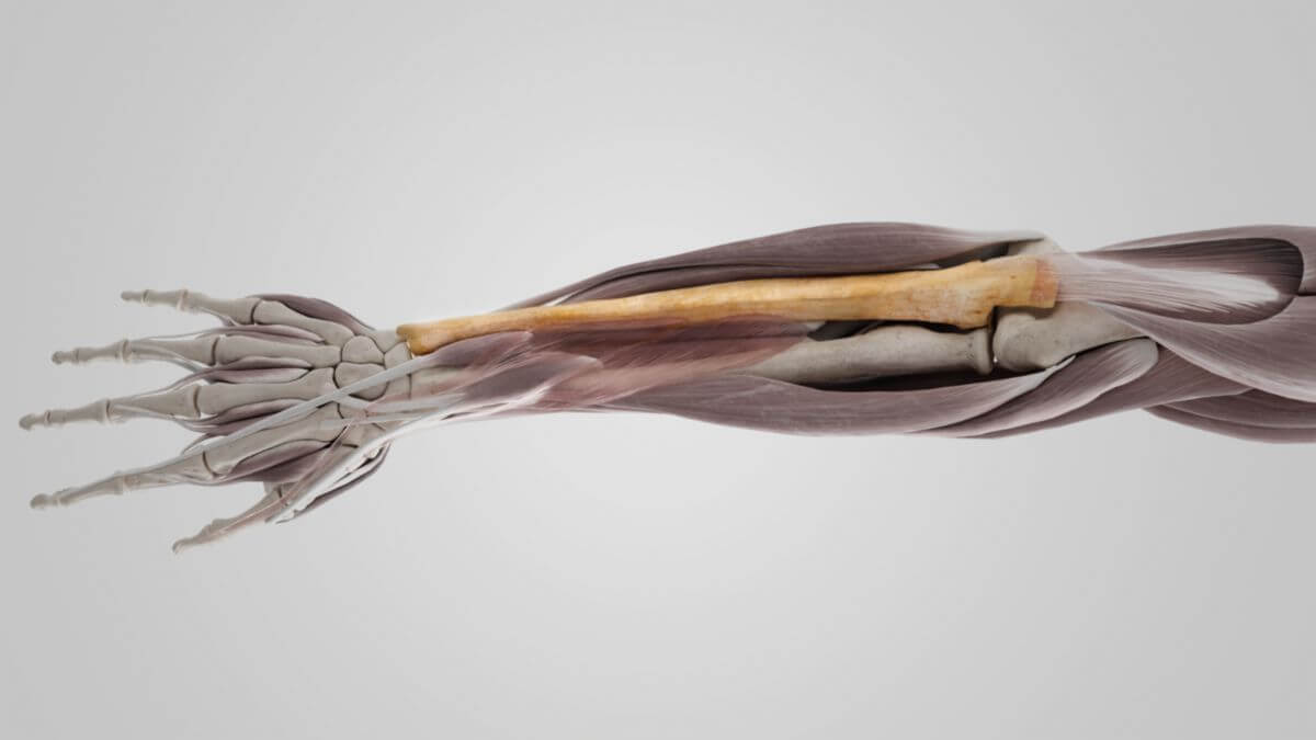
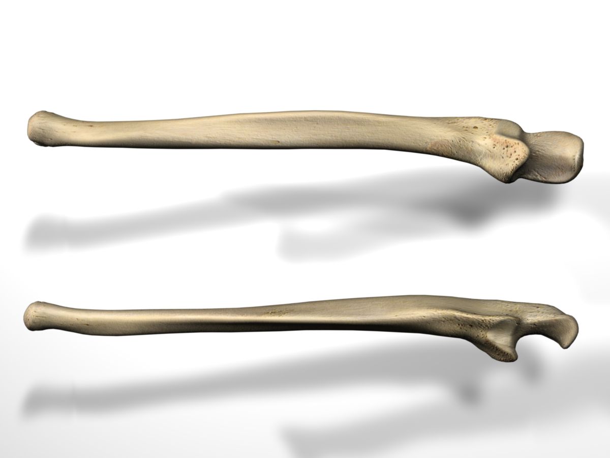
The ulna consists of a diamond-shaped body and a proximal and distal end piece. The strongly developed proximal end piece forms the largest part of the elbow joint. The smaller distal end piece is connected to the wrist via a cartilage disc.
2.1. Proximal end piece
Four important anatomical structures can be distinguished at the proximal end piece: the olecranon, the coronoid process, the trochlear incisura, and the radial incisura.
2.2. Olecranon
The olecranon is a prominent, beak-like bone projection located at the proximal end of the ulna. Its notable end extends into the olecranon fossa of the humerus, while its base narrows as it merges with the body of the ulna. The olecranon's posterior surface is broadly triangular and smooth, covered by the bursa of the elbow due to its direct subcutaneous position. Its superior surface, roughly diamond-shaped, is textured and serves as the attachment site for the triceps brachii muscle's tendon. A notable feature on the anterior edge of the olecranon is a transverse depression, which accommodates the posterior fibers of the elbow joint's capsule. In contrast, the anterior surface is smooth and concave, lined with articular cartilage, and forms the upper portion of the incisura trochlearis. The medial edge of the olecranon provides an origin point for some fibers of the flexor carpi ulnaris muscle, while the lateral edge is the attachment site for the anconeus muscle.
2.3. Coronoid process
The ulnar coronoid process, situated below the trochlear notch, is a prominent triangular bony extension. Its anteroinferior surface is notably concave and features a textured area, known as the ulnar tuberosity. This roughness is the point where tendon fibers of the brachialis muscle attach. On its lateral aspect, the coronoid process presents a trapezoidal, cartilage-coated articular surface, designed to accommodate the head of the radius. This specific area is known as the radial notch. Meanwhile, the medial side of this bony process serves as the origin point for two muscles: the flexor digitorum superficialis and the pronator teres muscle. During the action of flexion, the ulnar coronoid process interlocks with the coronoid fossa of the humerus, a key interaction for effective elbow joint movement.
2.4. Trochlear notch
The trochlear notch is a large, sickle-shaped indentation located between the olecranon and the coronoid process of the ulna. It is lined with articular cartilage, enabling smooth articulation with the trochlea of the humerus. This notch is characterized by a division into two parts: a lateral section and a slightly larger medial section, separated by a smooth bony ridge extending from the olecranon to the tip of the coronoid process.
2.5. Radial notch
Adjacent to the trochlear notch is the radial notch, a cartilage-lined indentation on the lateral aspect of the coronoid process. This structure is specifically shaped to accommodate the head of the radius. It features a slightly concave longitudinal profile, with its subtly raised edges providing an anchoring point for the annular ligament of the radius.
2.6. Shaft
The shaft of the ulna has an almost triangular cross-section in the proximal section with three edges and three surfaces separated by them. It becomes increasingly rounded distally.
2.7. Anterior margin
The anterior (volar) edge runs distally from the coronoid process. Its upper and middle section serves as the origin of the flexor digitorum profundus muscle. Fibers of the pronator quadratus muscle originate in the lower quarter. The margo volaris separates the anterior facies from the medial facies.
2.8. Posterior margin
The posterior (dorsal) edge of the ulna begins on the back of the olecranon and runs from there distally to the styloid process. It is very pronounced in the upper three quarters and serves as the base of an aponeurosis, which is the origin of the flexor carpi ulnaris muscle, the extensor carpi ulnaris muscle, and the flexor digitorum profundus muscle. The volar margin separates the posterior surface from the medial surface.
2.9. Interosseous margin
The medial edge is called the interosseous margin of ulna. It begins at the junction of two lines that extend distally from the edges of the radial surface. These lines enclose a triangular area of bone, the supinator crest, which marks part of the origin of the supinator muscle. The interosseous margin ends distally at the head of ulna. The edge is sharp-edged in the upper part and smooth and rounded in the lower section. The interosseous membrane, which connects the ulna to the radius, is attached to the interosseous membrane. It separates the posterior surface from the anterior surface.
2.10. Anterior surface
The anterior surface is significantly wider proximally than distally. The flexor digitorum profundus muscle originates in its upper 3/4. The lower quarter is covered by the pronator quadratus muscle.
2.10.1. Posterior surface
The posterior surface is broad and concave in the upper part, convex and somewhat narrower in the middle part and smooth and rounded in the distal part. A small bone edge runs diagonally across the upper part. The anconeus muscle inserts in the bone section above this edge. Below the bone edge, the surface is divided into two parts by a fine longitudinal conical ridge. The medial part is smooth and is covered by the extensor carpi ulnaris muscle. The lateral part is rough and serves – from top to bottom – as the origin of the following muscles:
- Supinator muscle
- Abductor pollicis longus muscle
- Extensor pollicis longus muscle
- extensor indicis muscle
2.10.2. Facies medialis
The facies medialis is the area of origin of the flexor digitorum profundus muscle in the proximal 3/4. The lower quarter borders on the integument.
2.10.3. Distal end piece (extremitas distalis)
The distal end of the ulna is much more delicate than the proximal end. Two prominent bone structures are found here.
2.10.4. Caput ulnae
The lateral caput ulnae is the distal joint head of the ulna. The distal part of the articular surface is in contact with the articular disc, which separates it from the proximal row of carpal bones. The lateral cartilage zone is taken up by the ulnar incisura of the radius.
2.10.5. Styloid process
Medial to the ulnar head is the styloid process of the ulna. It extends further distally than the head. Its rounded end serves as the attachment for the fibers of the ulnar collateral ligament of the wrist.
3. Topography
The ulna is used as a reference structure in the forearm. The term "ulnar" or "ulnaris" is used to denote a spatial reference to the ulna.
4. Development
The perichondral ossification of the ulna's body begins during the 7th embryonic week. The ossification centers in the distal epiphysis are laid down enchondrally between the 4th and 7th year of life, and in the styloid process between the 9th and 11th year of life. Proximally, the closure of the epiphyseal plate occurs earlier than distally.
5. Quiz
6. Image source
- Image source quiz: © DocCheck Flexikon