Corpus: Radius


1. Definition
2. Division
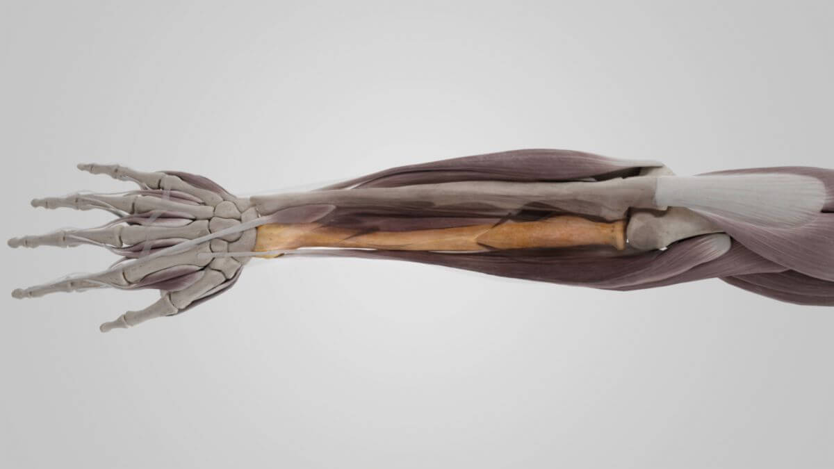
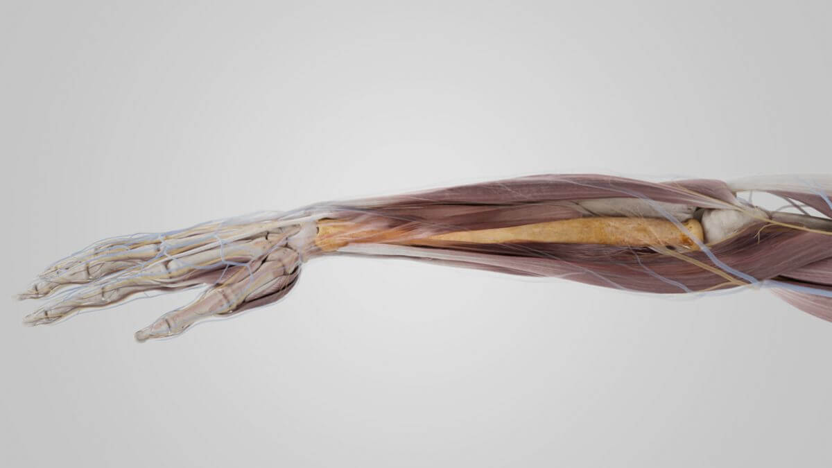
Like almost every other long bone, the radius can be divided into three sections:
- radial head: connects to the humerus
- radial neck
- radial shaft
While the radial head articulates with the head of the humerus, the so-called capitulum humeri, the distal end of the radius forms an articular surface for the carpal bones.
3. Surfaces
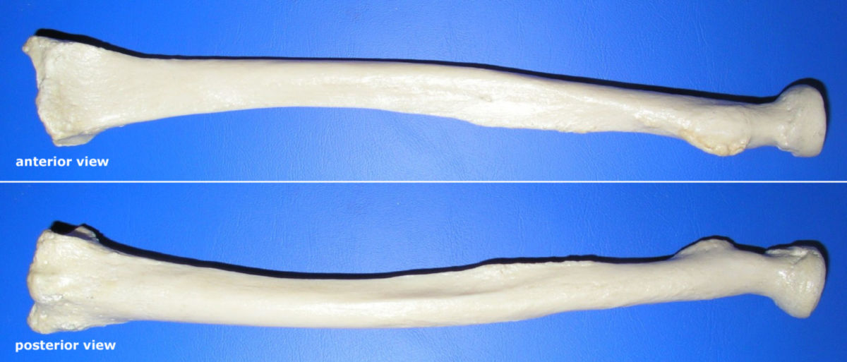
Despite its almost cylindrical body, various surfaces can be delineated on the radius due to the edges described above.
3.1. Anterior surface
The anterior surface is the bony surface located between the anterior margo and the interosseous margo. It serves as the point of origin for the middle and deep layer of the flexors of the forearm together with the interosseous membrane and the ulna.
3.2. Lateral surface
Between the margo anterior and the margo posterior there is also an area known as the facies lateralis. Some fiber tracts of the forearm muscles also originate here.
3.3. Posterior surface
This is the name given to the area that is spanned by the posterior margo and the interosseous margo. This also serves various forearm muscles, in this case the extensors, together with the interosseous membrane and the ulna as a surface of origin.
3.4. Carpal articular surface
The radius widens steadily distally so that its distal joint end has a cartilaginous articulation surface for the carpal bones, the carpal articular surface.
4. Noteworthy structures
4.1. Radial head
The cartilaginous head of the radius acts like a wheel that is placed on the neck of the radius. At its articulation surface with the humerus, it has an indentation for the humeral capitulum with which it forms a partial joint (articulatio humeroradialis) of the elbow joint (articulatio cubiti).
4.2. Radial tuberosity
A strong roughening of the radius is found slightly below the neck. This forms the attachment point of the main tendon of the biceps brachii muscle. During the pronation movement, the tendon is wound around the body of the radius, like a thread that is wound onto a spindle.
4.3. Styloid process of radius
If the radius is traced distally, a bony projection protrudes beyond the carpal articular surface at the lateral edge. This projection is known as the styloid process of the radius (processus styloideus radii). The brachioradialis muscle attaches to it.
4.4. Ulnar notch
The distal end of the radius has a notch on the medial side for the head of the ulna, the head of ulna. It is called the ulnar notch and forms the distal radioulnar articulation with the ulna.
5. Development
Perichondral ossification of the corpus radii begins during the 7th week of embryonic life. In contrast to the corpus, the epiphyses are formed enchondrally: the distal epiphysis at around 1 - 2 years of age, the styloid process at 10 - 12 years of age. The proximal epiphysis forms within the 4th to 7th year of life. The proximal epiphysis closes between the ages of 14 and 17, and the distal epiphysis does not close until the age of 20 (to 25).
6. Joints
The radius is involved in a total of 4 different partial joints. Firstly, it articulates with the humerus by means of the humeroradial articulation and secondly with the carpal bones by means of the radiocarpal articulation. The ulna has no direct involvement in the latter joint. It must bridge the distance resulting from the more proximal end of the joint with an articular disc. The ulna and radius also have two articular connections with each other. The proximal articulatio and the distal articulatio.
7. Quiz
8. Image source
- Image source quiz: © DocCheck Flexikon