Corpus: Biceps brachii muscle

Synonym: biceps muscle
1. Definition
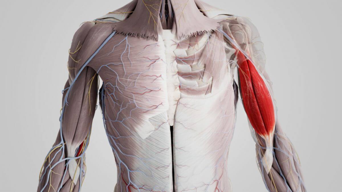
The biceps brachii muscle is a large two-headed and two-jointed muscle that belongs to the group of upper arm muscles. The two heads of the muscle are known as the long head and the short head. The muscle crosses two joints.
2. Anatomy
2.1. Origin
The long head originates from the supraglenoid tubercle of the scapula, while the short head arises sinewy from the coracoid process of the scapula.
2.2. Insertion
The biceps brachii muscle inserts at the radial tuberosity of the radius. Specifically, the short head inserts into the distal part of the tuberosity, while the tendon fibers of the long head extend more proximally into the apex of the tuberosity. The insertion tendon is partially or completely surrounded by the bicipitoradial bursa. In addition, an interosseous cubital bursa may be present in the direction of the ulna.
The bicapital aponeurosis, a superficial separation of the strong insertion tendon, inserts into the deep fascia of the forearm. The biceps brachii muscle thus crosses 2 joints, the shoulder and elbow joints.
2.3. Specific characteristics
The muscle is covered by the deltoid muscle. Therefore, the division can only be seen if this muscle is removed during dissection. The tendon of the longer head traverses the intertubercular sulcus of the humerus, passing through the joint capsule of the shoulder joint to its origin at the supraglenoid tubercle of the scapula. The tendon is surrounded by the intertubercular tendon sheath. The tendon's insertion in relation to the shoulder joint is intracapsular but extrasynovial.
The tendon at the origin of the short head is partially fused with the tendon at the origin of the coracobrachialis muscle.
2.4. Variety
In about 10 % an additional third head may arise from the humerus and join the muscle belly.
3. Innervation
The biceps brachii muscle is innervated by the musculocutaneous nerve from the brachial plexus (segments: C5-C6 or C7).
4. Topography
Two soft tissue grooves, the medial bicipital groove and the lateral bicipital groove, are located medially and laterally between the biceps and triceps muscles.
The medial bicipital groove contains the brachial artery, the ulnar nerve and the median nerve. The radial nerve traverses the lateral bicipital groove.
5. Function
5.1. Supination of the forearm
The biceps brachii muscle is the strongest supinator of the forearm, reinforced by the supinator muscle. Its supinator effect strengthens with increasing flexion and weakens when the forearm is extended - supination is achieved by the supinator muscle.
5.2. Flexion at the elbow joint
The biceps and the brachialis muscles are responsible for the flexion (bending) of the forearm at the elbow joint. The biceps is the direct antagonist of the triceps muscle. Flexion is strongest in the supination position and weak in the pronation position. The flexion effect of the brachioradialis and brachialis muscles is predominant in pronation.
5.3. Effect on the shoulder joint
In the shoulder joint, contraction of the long head causes a slight abduction movement of the arm and slight internal rotation, while contraction of the short head causes slight adduction. The contraction of both heads results in anteversion of the arm. It also stretches the forearm fascia via its insertion in the bicipital aponeurosis.
6. Clinic
The neurological examination of the innervation of the biceps brachii muscle is performed using the biceps reflex.
A traumatic rupture of the muscle tendon, known as a biceps tendon rupture, can occur, particularly in older people or because of overuse. The displacement of the long tendon of origin of the biceps brachii muscle from the intertubercular groove into the humeral joint due to a lesion of the biceps pulley is called a pulley lesion.