Corpus: Radial nerve


from Latin: radius - ray
Definition
Course
The radial nerve originates from the posterior cord of the brachial plexus. It contains nerve fibers from segments C5 to C8 and Th1. The radial nerve travels with the deep brachial artery through the slit of the triceps brachii muscle and from there continues its course into the canalis nervi radialis and musculospiral groove around the dorsal side of the humerus. It penetrates the lateral intermuscular brachial septum approximatly 10 cm proximal to the lateral epicondyle of the humerus. It joins the radial artery in the lateral bicipital sulcus and passes through the radial tunnel to the cubital fossa. There it gives off muscular branches to the brachioradial muscles and divides into a deep motor branch and a superficial sensory branch.
Deep branch
The deep motor branch runs on the dorsal side of the forearm through the supinator muscle and — by wrapping around the radius — reaches the extensor ligament of the forearm and from there to the hand. Its deep end branch is known as the posterior interosseous nerve.
Superficial branch
The superficial sensory branch is directed to the brachioradialis muscle and travels in the radial groove to the side of the radial artery. It passes under the brachioradialis tendon in the distal third of the forearm and thus reaches the extensor side. There it anastomoses with branches of the dorsal branch of the ulnar nerve. The superficial branch is divided into 5 dorsal digital nerves in its final segment.
Further branches
- posterior brachial cutaneous nerve: sensitively supplies the skin on the back of the upper arm
- dorsal antebrachial cutaneous nerve: begins in the radial groove, breaks through the superficial fascia, and finally supplies the skin on the extensor side of the forearm.
- posterior interosseous nerve: located on the interosseous membrane and supplies the deep layer of the extensor muscles of the forearm and the wrist.
- inferior lateral cutaneous nerve of the arm: provides sensory input to the lateral and dorsal skin of the upper arm.
Function
Motor innervation
The radial nerve primarily innervates the muscles on the dorsal side of the upper arm and forearm, including the extensors and the long muscles of the hand. It is therefore primarily responsible for the extension of the fingers and the hand.
Upper Arm
- triceps brachii muscle
- anconeus muscle
On the upper arm, a small part of the radial nerve also supplies the brachialis muscle — presumably with fibers involved in proprioception.
Forearm
- brachioradialis muscle
- extensor carpi radialis longus muscle
- extensor carpi radialis brevis muscle
- extensor indicis proprius muscle
- abductor pollicis longus muscle
- supinator muscle
- extensor digitorum muscle
- extensor pollicis longus muscle
- extensor pollicis brevis muscle
- extensor carpi ulnaris muscle
- extensor digiti minimi muscle
Sensory innervation
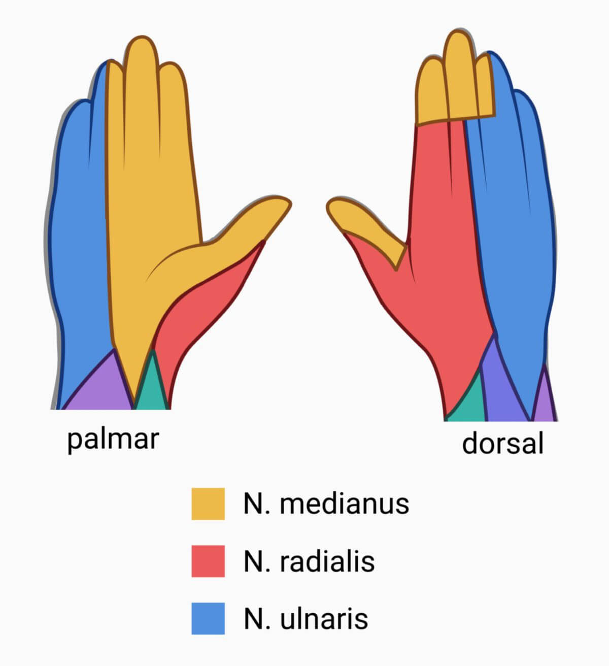
The radial nerve innervates the skin on the dorsal and lateral side of the arm. Its terminal branches supply the dorsal skin of the radial 2 1/2 fingers. The innervation of the two end phalanges of fingers I to III of the other hand, is largely supplied by the digital palmar nerves of the median nerve.
Clinic
A lesion of the radial nerve is referred to as radial nerve palsy. A distinction is made based on the localization of the lesion:
- High radial nerve palsy
- Middle radial nerve palsy
- Low radial nerve palsy
In high radial nerve palsy, for example after a humerus fracture, the entire extensor muscles of the arm and hand become paralyzed. The most common symptom is a flaccid drooping of the hand at the wrist (drop hand). The triceps tendon reflex and the radial periosteal reflex are weakened. Lower radial nerve palsy is caused by compression of the radial nerve in the supinator tunnel, also known as supinator tunnel syndrome. Injury to the sensitive superficial branch leads to Wartenberg's syndrome.