Corpus: Glenohumeral joint

Synonym: shoulder joint
1. Definition
2. Structure
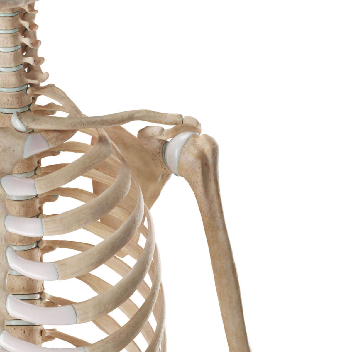
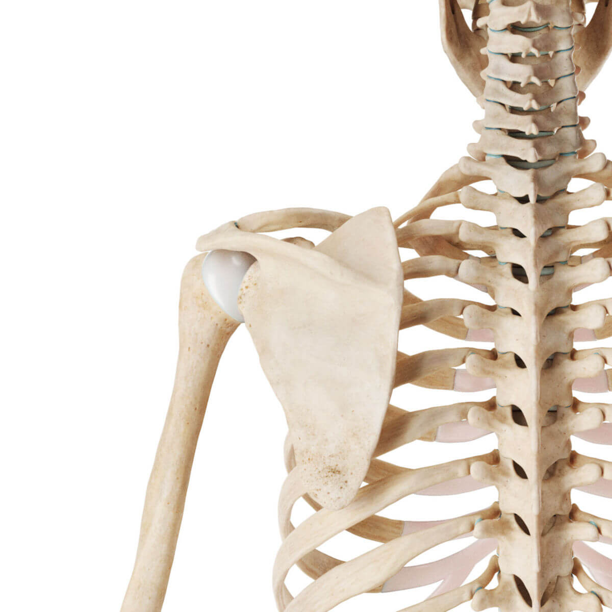
The spherical head of the humerus articulates with the longitudinally oval-shaped glenoid cavity, or "glenoid." It is relatively small compared to the humeral head and therefore does not completely enclose it, as seen in the hip joint, for example. An increase in the contact area between both joint partners is achieved by the 3 to 4 mm wide labrum glenoidale (glenoid labrum) formed around the glenoid cavity. The labrum glenoidale consists of fibrocartilage and is attached to the glenoid cavity.
The glenohumeral joint capsule is relatively spacious, thin, and lax. Caudally, in a relaxed position, there is a reserve zone of about 1 cm in length, known as the axillary recess. If the arm is immobilized in this position for too long, this lower capsule fold shrinks, leading to movement restrictions. For this reason, during prolonged immobilization, the glenohumeral joint is fixed in an abducted and slightly anteriorly rotated position.
The capsule extends cranially to the coracoid process and includes the tendon of the long head of the biceps brachii muscle, which is anchored to the glenoid labrum and the supraglenoid tubercle. The supraglenoid and infraglenoid tubercles of the scapula are integrated into the joint capsule and thus not palpable from the outside. In contrast, the two tubercles of the humerus are located outside the capsule.
3. Ligaments
The glenohumeral joint has a relatively weakly developed ligamentous apparatus in relation to its demands. Guidance by ligaments is only provided to a limited extent. The main ligaments are:
These ligaments reinforce the glenohumeral joint cranially and ventrally. There are no relevant fiber bundles dorsally. Traumatic shoulder dislocation usually occurs anteriorly and inferiorly.
Adjacent ligaments without direct influence on the glenohumeral joint include the transverse humeral ligament, which holds the biceps tendon in the intertubercular groove of the humerus, and the coracoacromial ligament.
4. Muscles
The guidance and stabilization of the glenohumeral joint are provided by muscles that encircle it like a cuff — the so-called rotator cuff. It contributes significantly more to stability than the ligaments. However, the muscle cuff is not completely closed but has gaps. One of these gaps is the triangular rotator interval.
5. Bursae
Numerous bursae play an important role in the function of the glenohumeral joint.
The subscapular bursa reduces friction between the tendon of the subscapular muscle and the scapula when lying beneath the tendon. It communicates with the joint cavity through an oval opening. The subcoracoid bursa is a reserve space of the joint located below the coracoid process. It also communicates with the joint cavity.
The subacromial and subdeltoid bursae are also referred to as the subacromial joint. However, anatomically, the term is not correct as there are no cartilage-covered joint surfaces, and therefore, the prerequisites of a joint are not met. These bursae ensure the mobility of the greater tubercle of the humerus under the acromion during arm abduction.
6. Situs
7. Mechanics
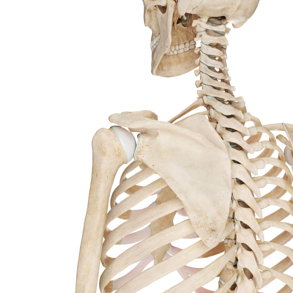
Due to its ball-and-socket structure, movement of the arm is possible in all three axes. The accessory joints of the shoulder girdle (acromioclavicular joint, sternoclavicular joint) make a significant contribution to mobility. Depending on the requirements, the positions of the clavicle and scapula are modified by the mobility of these joints.
In practice, it should be assumed that hardly any arm movement can be attributed solely to movement of the glenohumeral joint. Therefore, the interaction of the joint group must be taken into account when assessing movement restrictions.
The ranges of motion in different axes are indicated below, starting from the neutral position:
- Transverse
- Anteversion up to 90° in the glenohumeral joint, with elevation beyond (up to a maximum of 170°) involving the joints of the shoulder girdle; complete elevation (180°) is possible by extending the spine
- Retroversion up to a maximum of 50°
- Sagittal
- Abduction up to 90° in the glenohumeral joint, with involvement of the shoulder girdle and spine up to 180°
- Adduction up to 45°
- Vertical Axis
- Internal rotation up to 30° with the upper arm close to the body
- External rotation up to 60° with the upper arm close to the body
8. Clinic
8.1. Diseases
Important diseases or injuries of the glenohumeral joint include:
- Shoulder dislocation
- glenohumeral joint arthritis (omarthrosis)
- Frozen shoulder
- Bankart lesion
- SLAP lesion
- Hill-Sachs lesion
8.2. Diagnostics
Several clinical tests are available to examine the glenohumeral joint, including:
- Global tests: e.g., neck grip, apron grip
- Specific tests: e.g., infraspinatus test, Jobe test, belly press test
Advanced diagnostic techniques include imaging (X-ray, MRI) and glenohumeral joint arthroscopy. Standard X-ray images include:
- Shoulder AP
- Axial shoulder
For specific questions, an MRI of the shoulder is performed.