Corpus: Clavicle


Synonym: collar bone
1. Definition
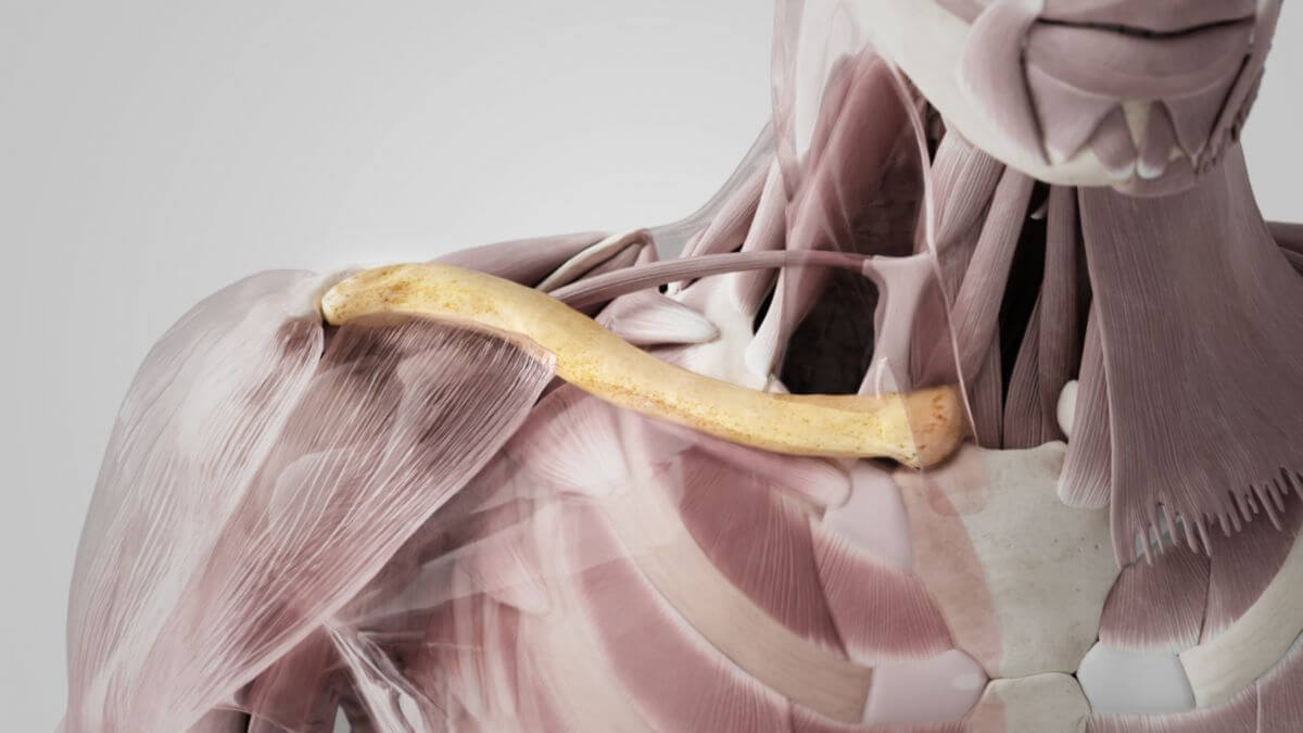
The clavicle is the s-shaped curved bone whose terminal articular surfaces are connected medially to the sternum and laterally to the scapula and is thus involved in the movement of the shoulder joint.
2. Anatomy
The clavicle forms the anterior part of the bony shoulder girdle. It is positioned almost horizontally above the anterior upper part of the thorax. The bone consists of a shaft (corpus claviculae) and two ends:
- the medially located sternal end and
- the lateral acromial end
2.1. Corpus claviculae
Due to its cross-section, the shaft of clavicle can be divided into a flattened lateral third and the round or prismatic medial two thirds.
2.2. Lateral third
The superior aspect of the lateral third of the clavicle is flattened and rough. Anteriorly, it shows depressions into which the fibers of the deltoid muscle, which has part of its origin here, radiate. Posteriorly, corresponding structures can be seen through the insertion of the trapezius muscle. The intervening part of the bone borders directly on the subcutaneous tissue and is palpable under the skin. The inferior aspect of the lateral third is flat. It shows two interesting structures:
- the conoid tubercle, into which the conoid ligament radiates and
- the trapezoid line, to which the trapezoidal ligament attaches.
2.3. Medial two-thirds
The medial two-thirds of the clavicle have a triangular cross-section. They curve convexly anteriorly and concavely posteriorly. They have three edges (margo superior, margo anterior and margo posterior) and three surfaces (facies): The anterior surface (facies anterior) lies between the margo superior and anterior. It is located almost below the surface of the skin and is covered by the platysma. Fibers of the pectoralis major muscle originate at its lower end and the sternocleidomastoid muscle at its upper end. The posterior surface (posterior facies) is topographically related to the brachial plexus and the subclavian artery and vein. Fibers of the sternohyoid muscle originate near the extremitas sternalis. The lower surface (inferior facies) has a broad, rough surface in its medial area, the costal tuberosity, to which the costoclavicular ligament attaches. The rest of the surface is filled by a bone depression that serves as the attachment of the subclavian muscle. The coracoclavicular fascia attaches to the edges of the hollow.
2.4. Joints
The clavicle is connected to the neighboring bones via two joints:
- the sternal end with the manubrium of the sternum via the sternoclavicular joint
- the acromial end with the acromion of the scapula via the acromioclavicular joint
3. Development
The clavicle is the first ossifying skeletal element of the body. Its diaphysis is already formed in the 6th to 7th embryonic week through desmal ossification. Only the epiphyses have a cartilage core (chondral ossification). Ossification of the sternal end begins in the 18th to 20th year of life, the closure of the epiphyseal groove does not take place until the 20th to 24th year of life. Since desmal ossification presumably originates from two separate ossification centers, subsequent disruption of their fusion can lead to congenital clavicular pseudarthrosis. Hypoplasia or aplasia of the clavicle occurs in various malformation syndromes, e.g. in kleidocranial dysplasia or mandibuloacral dysplasia.
4. Topography
The line running through the center of the clavicle is called the medioclavicular line.
5. Clinic
The clavicle and its joints are frequently affected by injuries due to their exposed position. These include clavicle fracture and acromioclavicular joint dislocation. The partial or complete surgical removal of the clavicle is called claviculectomy.