Corpus: Sternocleidomastoideus muscle


from Latin: sternum - breastbone
from ancient Greek: κλείς ("kleis") - key; μαστός ("mastos") - breast
1. Definition
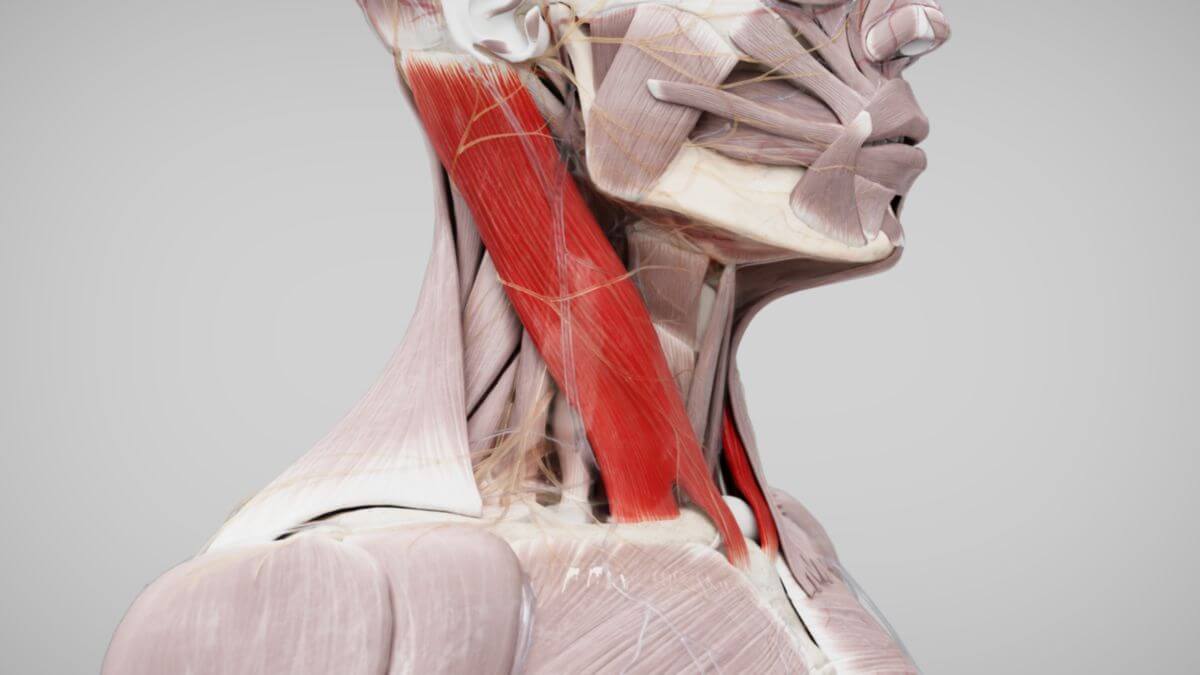
The sternocleidomastoid muscle belongs to the superficial layer of the ventral neck musculature. It is a two-headed muscle with a lateral head (caput laterale) and a medial head (caput mediale).
2. Course
2.1. Origin
The lateral head (caput laterale) originates from the upper edge and anterior surface of the medial third of the clavicle (collarbone), with its fleshy and aponeurotic fibers running almost vertically upward. The medial head (caput mediale) originates from the anterior surface of the manumbrium of the sternum, with its fibers running cranially, laterally, and dorsally.
The origins of both muscle heads form a small triangular area with the clavicle, known as the lesser supraclavicular triangle, which appears on the surface of the skin as the minor supraclavicular fossa. The two heads unite approximately in the middle of the neck to form a thick, rounded muscle belly.
2.2. Attachment
The sternocleidomastoid muscle primarily inserts on the lateral side of the mastoid process of the temporal bone. It also attaches to the lateral half of the superior nuchal line of the occipital bone via a thin aponeurosis.
3. Topography
The sternocleidomastoid muscle divides the lateral part of the neck into two triangles:
- lateral triangle of the neck
- anterior triangle of the neck
When viewed from the front, the right and left sternocleidomastoid muscles form a characteristic "V" shape in the muscle contour of the neck.
The region overlying the muscle itself is called the sternocleidomastoid region. Beneath the muscle, the common carotid artery, internal jugular vein, vagus nerve, and cervical artery run together as a vascular-nerve bundle. Sensory nerve branches from the cervical plexus emerge posterior to the sternocleidomastoid muscle at a point known as Erb's point.
4. Innervation
The sternocleidomastoid muscle receives motor innervation from the accessory nerve (cranial nerve XI) and direct nerve branches from the cervical plexus, specifically from segments C1 to C3/C4.
5. Function
Unilateral contraction of the sternocleidomastoid muscle results in lateral flexion of the head toward the shoulder and a slight backward flexion, with simultaneous rotation of the head to the opposite side. Bilateral contraction causes dorsiflexion (extension) of the head. When the head is fixed, both muscles work together as auxiliary respiratory muscles.
6. Clinic
Congenital malformations of the sternocleidomastoid muscle, leading to unilateral shortening, can result in congenital muscular toricollis. The carotid pulse can be palpated at the anterior edge of the middle third of the muscle. Additionally, the medial edge of the sternocleidomastoid muscle serves as an important landmark when inserting a central venous catheter into the internal jugular vein.