Corpus: Pectoralis major muscle


from Latin: pectus - chest
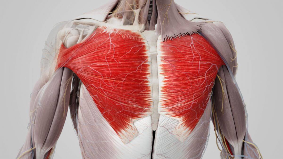
1. Definition
The pectoralis major muscle is a large, strong, and fan-shaped muscle located on the upper part of the bony thorax, directly above the pectoralis minor muscle. It is one of the outer pectoral muscles that connect the ventral shoulder girdle to the trunk. The muscle is divided into three parts:
- Clavicular part
- Sternocostal part
- Abdominal part
2. Progression
The three parts of the pectoralis major muscle have distinct origins:
- The clavicular part originates from the medial half of the clavicle
- The sternocostal part arises from the ipsilateral edge of the sternum, specifically the manubrium and body of the sternum, as well as the cartilages of the second to sixth (sometimes seventh) ribs
- The abdominal part originates from the anterior leaf of the aponeurosis of the rectus abdominis muscle
The muscle fibers converge concentrically towards their insertion, forming a flat tendon that attaches to the crest of the greater tubercle of the humerus. This tendon consists of two superimposed layers. The anterior layer, which is thicker, is formed from the fibers of the clavicular and upper sternal parts. It inserts at the crest in the same order as the muscle origin, with the clavicular part inserting most cranially and the sternal fibers inserting more caudally. The posterior layer receives the majority of the sternal muscle fibers and deep fibers from the ribs, running behind the anterior layer along the humerus and inserting higher up, creating a twisted appearance. This twisting is undone with the elevation of the arm, forming a small niche on the dorsal surface of the muscle, known as the pectoralis pouch.
3. Topography
The pectoralis major muscle lies beneath the pectoralis fascia, to which it is firmly attached, and above the clavipectoralis fascia. The infraclavicular fossa (or "Mohrenheim pit") is located between the clavicular part of the pectoralis major muscle, the deltoid muscle, and the clavicle, serving as a passageway for the cephalic vein, which pierces the clavipectoral fascia.
4. Innervation
The pectoralis major muscle is innervated by the medial pectoral nerve (C8-Th1) and the lateral pectoral nerve (C5-C7) from the brachial plexus, specifically from the lateral and medial fasciculi.
5. Blood supply
Arterial blood is supplied to the pectoralis major muscle by muscular branches of the superior thoracic artery and the pectoral branch of the thoracoacromial artery. Venous blood is drained through the pectoral veins into the subclavian vein.
6. Varieties
The area of origin and distribution of the pectoralis major muscle can vary, especially in its attachment sites on the sternum and ribs. Parts of the muscle, such as the sternocostal or abdominal parts, may be absent. A complete absence of the muscle occurs in Poland syndrome. Another variation includes partial fusion of the muscle with the deltoid or latissimus dorsi muscles.
7. Function
The pectoralis major muscle is responsible for anteversion, adduction, and internal rotation of the arm at the shoulder joint. When the arms are fixed, as in a propped-up position (often referred to as the "coachman's seat"), the pectoralis major serves as a strong inspiratory muscle, aiding in breathing, particularly during strenuous activities, alongside the pectoralis minor muscle.
8. Clinic
Injuries to the pectoralis major muscle are rare and typically occur in young men aged 20 to 40, often associated with weightlifting and other sports activities.