Corpus: Latissimus dorsi muscle


1. Definition
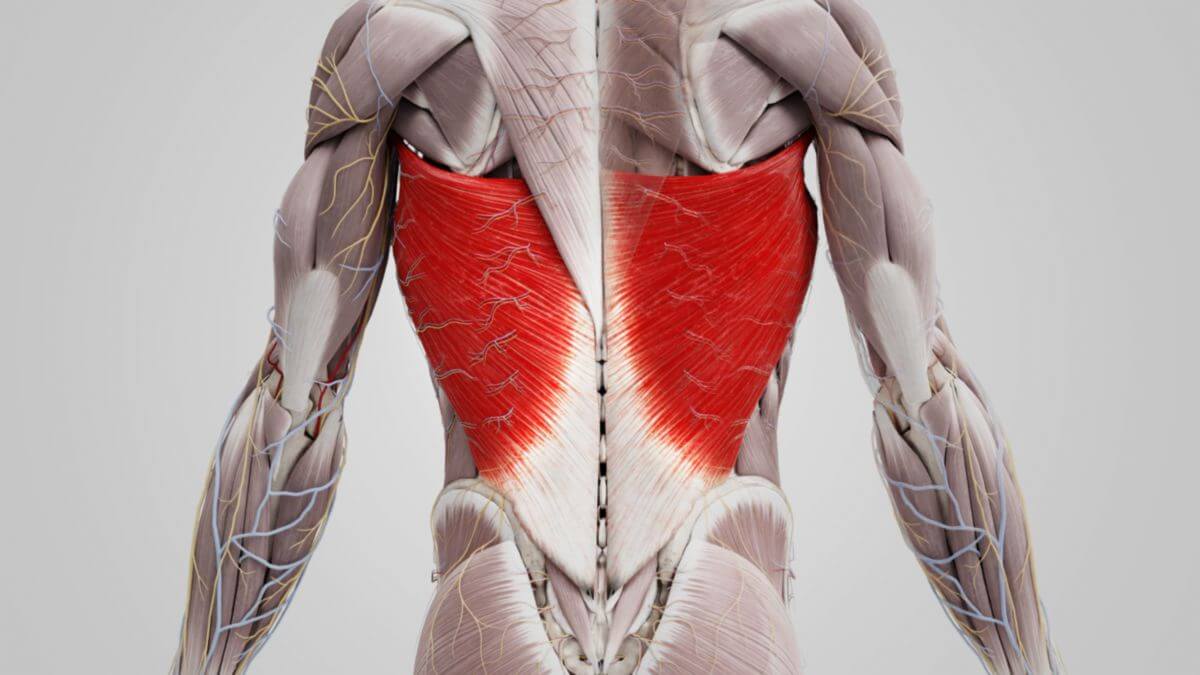
The latissimus dorsi muscle is a large, superficial skeletal muscle located on the back. It is part of the secondary back musculature, influencing the shoulder girdle from the dorsal side. In terms of surface area, the latissimus dorsi is the largest skeletal muscle in the human body.
2. Course
2.1. Origin
The latissimus dorsi muscle has a broad area of origin, which includes the following points:
- Spinous processes of thoracic vertebrae T7-T12 and lumbar vertebrae L1-L5
- Sacrum
- Supraspinous ligament and thoracolumbar fascia
- 9th to 12th ribs
- Crista iliaca of the iliac bone
The extent of the origin area can vary significantly from person to person. The muscle is typically divided into three anatomical parts based on its origin zones:
- Vertebral part
- Costal part
- Iliac part
Additionally, there is sometimes an inconstant origin at the inferior angle of the scapula, referred to as the scapular part. The muscle fibers converge from these origin areas, extending cranially and laterally toward their insertion on the upper arm. The latissimus dorsi muscle, along with the teres major muscle, forms the posterior axillary fold.
2.2. Attachment
The latissimus dorsi muscle attaches to the crest of the lesser tubercle and the intertubercular sulcus of the humerus, situated between the insertions of the teres major and pectoralis major muscles. This attachment is localized on the medial side of the humeral head.
At the junction with the teres major muscle lies the subtendinous bursa of latissimus dorsi muscle, which can become inflamed and cause pain, a condition known as bursitis.
3. Innervation
The latissimus dorsi muscle is innervated by the thoracodorsal nerve, which originates from the infraclavicular part of the brachial plexus, specifically from spinal cord segments C6-C8.[1]
4. Topography
The anterior border of the latissimus dorsi muscle, together with the posterior border of the obliquus externus abdominis muscle and the crista iliaca, forms the lumbar triangle.
5. Varieties
A variant of the latissimus dorsi muscle includes the presence of aberrant muscle fibers extending to the pectoralis major muscle, known as the muscular axillary arch. This arch can be 7 to 10 cm long and 5 to 15 mm wide, and as it crosses the axillary pathways, it may cause impingement.
6. Function
The latissimus dorsi muscle plays a crucial role in several functions. It is responsible for adduction and internal rotation of the arm, bringing the arm closer to the body and rotating it inward. The muscle also supports retroversion, aiding in moving the arm backward. Additionally, it acts as an antagonist to the deltoid muscle, counteracting its actions. When the arm is fixed, the latissimus dorsi can pull the upper body toward the arm, making it essential for climbing. During expiration, particularly when coughing, the latissimus dorsi functions as an accessory respiratory muscle, which can lead to muscle soreness after prolonged coughing.
7. Clinic
The latissimus dorsi muscle, together with the trapezius muscle and the medial edge of the scapula, forms a muscle-weak area that can be used for auscultation of the lungs when the scapula is moved ventrolaterally. To facilitate this, the patient is asked to cross their arms in front of their chest.
Surgically, the latissimus dorsi muscle is used in breast reconstruction following mastectomy. It has also been historically used in treating advanced heart failure as part of a procedure known as cardiomyoplasty.
8. Source
- ↑ Trepel, M: Neuroanatomie: Struktur und Funktion p. 36