Corpus: Skeletal musculature


Synonym: skeletal muscles
1. Definition
The skeletal musculature includes muscles, which are attached to the skeleton or – in a broader sense – are responsible for body movement. They belong to the striated muscle group, similar to cardiac muscle.
Note: The term "skeletal muscle" can be misleading, as it includes muscles not directly connected to the skeleton, such as tongue muscles, laryngeal muscles, and parts of the facial muscles.
2. Anatomy
2.1. Macroscopic anatomy
Skeletal muscles are broadly divided into a muscle head and a muscle belly. Based on the number of muscle heads, they can be categorized as:
- Single-headed (e.g., brachialis muscle)
- Two-headed (e.g., biceps brachii muscle)
- Three-headed (e.g., triceps brachii muscle)
- Four-headed (e.g., quadriceps femoris muscle)
Each muscle has points of origin and insertion, connected to bones by tendons or directly. A single skeletal muscle may have multiple points of origin, even on different bones.
Skeletal muscles also exhibit different types of pinnation, categorized into single-pennate and double-pennate muscles, based on the arrangement of fibers relative to the central tendon.
2.2. Functional anatomy
Skeletal muscles are categorized based on their interaction and biomechanical roles:
- Agonist
- Antagonist
- Synergist
Skeletal muscles that act in the same direction, i.e. complement each other in their effect, are called agonists. If they act in opposite directions, they are called antagonists.
Based on their primary movement, skeletal muscles are further classified as:
- Flexors muscles
- Extensors muscles
- Rotators: skeletal muscles that generate a rotational movement
- Adductors: skeletal muscles that pull a limb towards the body
- Abductors: skeletal muscles that move a limb away from the body
2.3. Systematics
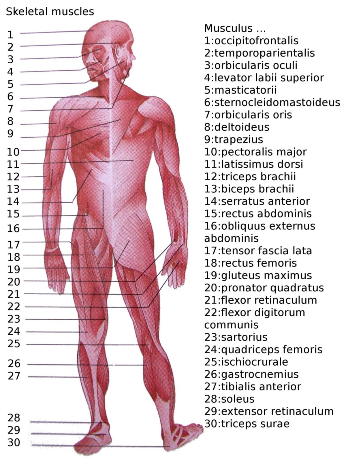
To facilitate understanding, skeletal muscles are grouped based on their location and function, such as upper limb, lower limb, trunk, and head/neck muscles.
see also: Human skeletal musculature
2.4. Histology
A skeletal muscle is composed of contractile muscle fibers and auxiliary connective tissue. Muscle fibers form a functional syncytium, meaning they operate as a unified structure rather than independent cells.
The skeletal muscle is surrounded externally by a thick layer of connective tissue called the fascia. From this outer layer, connective tissue extensions penetrate into the muscle, dividing it into smaller groups of fibers known as septa. These divisions create a hierarchical structure within the muscle, with the sarcomere being the smallest functional unit responsible for muscle contraction.
The skeletal muscle structure can be divided into hierarchically organized units:
| Plane | Unit | Surrounding structure |
|---|---|---|
| 1 | Muscle | Fascia, epimysium |
| 2 | Bundle of muscle fibers | Perimysium |
| 3 | Muscle fiber | Endomysium |
| 4 | Myofibril | Basallamina |
| 5 | Sarcomere | Basallamina |
| 6 | Myofilaments |
The connective tissue layers surrounding the muscle fibers and fiber bundles converge at the muscle ends to form tendons, which attach the muscle to bone. These connective tissues also house the nerves and blood vessels that supply the muscle with signals and nutrients.
The primary components of skeletal muscle are the contractile proteins:
- Actin, which makes up about 3 % of the total muscle weight.
- Myosin, which accounts for 7 % of the total muscle weight.
These proteins are organized within sarcomeres, the functional units of muscle contraction, defined as the distance between adjacent Z-discs where actin filaments are anchored. Contractile proteins make up approximately 100 mg per gram of skeletal muscle tissue.
Skeletal muscle regeneration relies on satellite cells, specialized stem cells that facilitate repair and growth of damaged muscle fibers.
3. Physiology
Skeletal muscles are essential for maintaining body posture and enabling movement, forming the majority of the muscle mass in the human body.
Their function is explained by the sliding filament theory, which describes how actin and myosin filaments slide past each other to produce contraction. Skeletal muscles are controlled by motor nerves that transmit electrical impulses to the motor end plate through the release of acetylcholine, initiating muscle activity. This activity is regulated both voluntarily, through signals from the motor cortex, and involuntarily, via spinal reflexes. The length of skeletal muscles is adjusted to the position of the joints through a length control system, ensuring smooth and coordinated movements.
4. Biochemistry
Skeletal muscle cells produce and secrete signaling molecules called myokines.