Corpus: Sacrum


from Latin: sacer - sacred
1. Definition
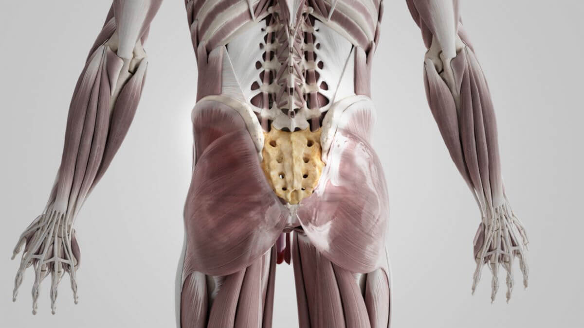
The sacrum is a wedge-shaped bone composed of five fused vertebrae, known as sacral vertebrae. It is a part of the human spine and forms the posterior section of the bony pelvis.
2. Anatomy
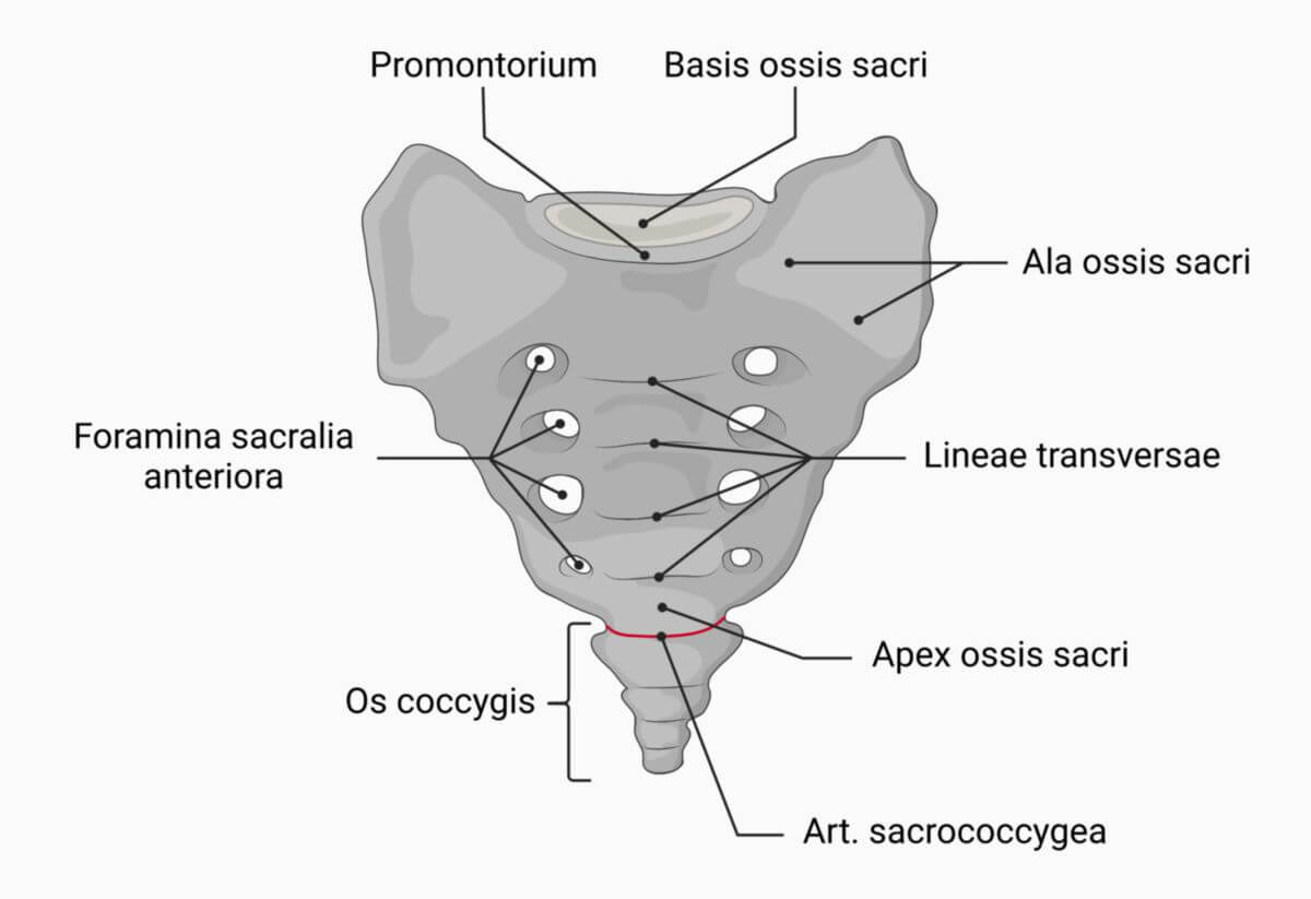
The sacrum has a blunt upper end, known as the base, a pointed lower end called the apex, and three distinct surfaces: the anterior surface (pelvic surface), the posterior surface (dorsal surface), and the lateral surfaces. The base of the sacrum is the broad upper part that connects to the last lumbar vertebra (L5) through an intervertebral disc. The most ventrally projecting point of the base is known as the promontory. The apex of the sacrum is the caudal end that connects to the coccyx.
A distinctive feature of the sacrum is the presence of four pairs of openings on both its anterior and posterior surfaces:
- Anterior sacral foramina
- Posterior sacral foramina
2.1. Pelvic surface
The pelvic surface is the anterior concave surface of the sacrum. It features fine transverse ridges, called transverse lines, which represent the original intervertebral spaces. On either side of these ridges are four roundish openings, the foramina sacralia anteriora. These foramina, which decrease in size from cranial to caudal, allow the anterior rami of the sacral nerves to exit and the lateral sacral arteries to enter.
The lateral part of the sacrum surrounding these foramina is known as the lateral part. The upper portion of this region forms the sacral wing, which is part of the terminal line. The piriformis muscle originates from the lateral surface near the second to fourth sacral foramina.
2.2. Dorsal surface
The dorsal surface is the posterior, convex surface of the sacrum, featuring one unpaired and two paired bony ridges formed by the fusion of the spinous, articular, and transverse processes of the sacral vertebrae:
- Median sacral crest
- Intermediate sacral crest
- Lateral sacral crest
The multifidus muscle originates on either side of the prominent median sacral crest in a shallow bony groove formed by the fused laminae of the sacral vertebrae. Incomplete fusion of the laminae of the fourth and fifth sacral vertebrae creates the sacral hiatus, an opening in the lower part of the sacral canal.
The sacral canal, which continues from the spinal canal, has a triangular cross-section above the sacral hiatus. It houses the bundled sacral nerves, which exit through the anterior and posterior sacral foramina.
The intermediate sacral crest is formed by the fused articular processes, with the superior articular process of the first sacral vertebra at its upper end. This process articulates with the inferior articular process of the fifth lumbar vertebra. The inferior articular process of the sacrum articulates with the cornua of the coccyx.
Laterally, the four foramina sacralia posteriora allow the posterior rami of the sacral nerves to pass through. These foramina are smaller and more irregular in shape compared to the anterior foramina.
The lateral sacral crest is formed by the fused transverse processes, which extend laterally from the posterior sacral foramina. These transverse tuberosities serve as attachment points for ligaments, such as the posterior sacroiliac ligament and the sacrotuberous ligament.
2.3. Lateral surface
The lateral surface begins broadly at the cranial end and narrows towards the caudal end. The upper half features an ear-shaped surface, the facies auricularis, which articulates with the ilium of the pelvis. This surface is covered with cartilage during adolescence. Behind the auricular surface lies the sacral tuberosity, a rough bony area where the posterior sacroiliac ligaments attach.
The lower half of the lateral surface is thin and serves as an attachment point for the sacrotuberous and sacrospinous ligaments, as well as fibers of the gluteus maximus muscle. It ends caudally in a bony projection called the inferior lateral angle. Medial to this angle is a notch that forms a foramen with the transverse process of the first coccygeal vertebra, allowing the passage of the anterior branch of the fifth sacral nerve.
2.4. Joints
The sacrum is involved in three major joints:
2.4.1. Lumbosacral joint
The lumbosacral joint, located between the last lumbar vertebra (L5) and the base of the sacrum. This compound joint includes the intervertebral disc between L5 and the sacral base and the facet joints between the superior articular processes of the sacrum and the inferior articular processes of L5.
2.4.2. Sacrococcygeal joint
The sacrococcygeal joint, located at the caudal end of the sacrum, where the apex of the sacrum articulates with the cranial surface of the coccyx. This joint can be a true synovial joint or a synchondrosis.
2.4.3. Sacroiliac joint
The sacroiliac joint, where the sacrum articulates with the ilium of the pelvis via the ear-shaped auricular surface on both sides.
3. Biomechanics
The sacroiliac joint has limited mobility due to the strong ligaments that bind the bones. The primary movement allowed is called nutation, where the base of the sacrum tilts ventrally around a transverse axis through the second sacral vertebra. The opposite movement, where the sacral base tilts dorsally, is called counternutation.
4. Clinic
Due to its robust structure and large bone mass, the sacrum is resistant to fractures. However, isolated sacral fractures are rare and typically occur only with significant trauma, often as part of complex pelvic fractures.