Corpus: Sphenoid bone


1. Definition
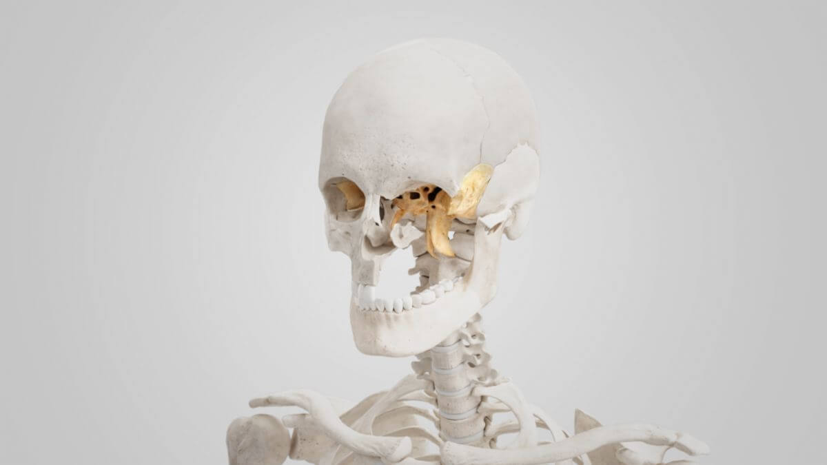
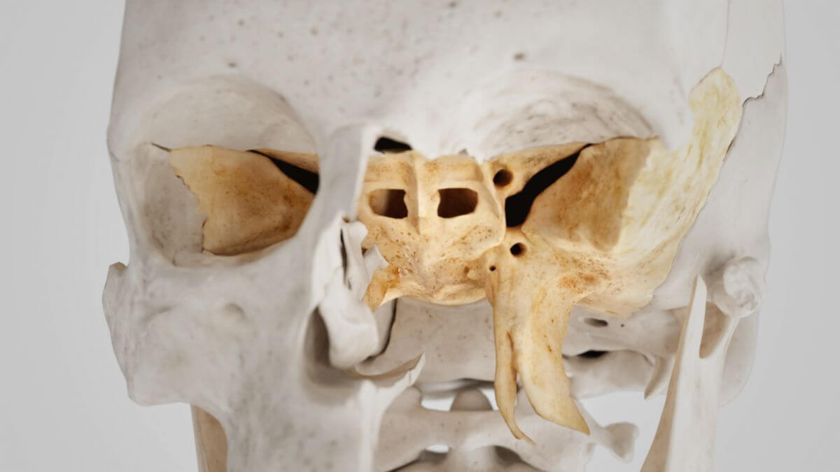
The sphenoid bone is a cranial bone of the neurocranium, located in front of the temporal and the occipital bones at the base of the skull. Viewed dorsally, it resembles a butterfly with a body, two paired laterally extending wings (greater and lesser wings) and caudally extending pterygoid processes.
2. Body of the sphenoid bone
The body of the sphenoid bone is roughly square shaped. Inside are two cavities, the sphenoid sinuses, separated by a septum.
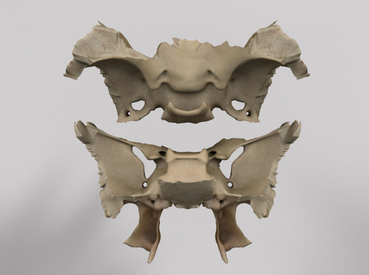
2.1. Superior surface
From a ventral to dorsal view, the superior surface of the sphenoid body reveals several important anatomical structures:
- The ethmoidal spine articulates with the ethmoid bone
- The jugum sphenoidale connects the two small wings of the sphenoid bone
- The chiasmatic sulcus: contains the optic chiasm
- The optic foramen is located lateral to the chiasmatic sulcus on both sides, allowing passage for the optic nerve and the ophthalmic artery into the skull.
- The tuberculum sellae and processus clinoideus medius are located on both sides
- The sella turcica is the deepest part, the hypophyseal fossa, houses the pituitary gland
- The dorsum sellae ends on both sides in a posterior clinoid process
- The notch is located lateral to the dorsum sellae, receiving the abducens nerve
- The petrosal process articulates with the petrous part of the temporal bone
- The clivus is a shallow pit that slopes dorsally and caudally towards the foramen magnum, merging with the cranial surface of the neighboring occipital bone
2.2. Lateral surface
The lateral surfaces of the sphenoid body give rise to the two large sphenoid wings. Directly above their attachment is an S-shaped groove (carotid sulcus) on both sides, which accommodates the internal carotid artery and the cavernous sinus. This groove is separated from the wing by a bony edge, the lingula.
2.3. Posterior surface
The posterior surface of the sphenoid body articulates with the occipital bone. In childhood, this connection is made by a cartilaginous plate that ossifies between the age of 18 and 25.
2.4. Anterior surface
The anterior surface of the sphenoid body features a central bony crest, the sphenoidal crest, which articulates with the perpendicular plate of the ethmoid bone, forming part of the nasal septum. Other parts of the anterior surface articulate with the ethmoid bone, frontal bone, and palatine bone. Irregular openings lead into the sphenoid sinuses on both sides, which communicate with the upper and posterior part of the nasal cavity and are partially closed by the sphenoidal conchae.
2.5. Inferior surface
A triangular bony ridge, the sphenoidal rostrum, protrudes from the lower surface of the sphenoid body. It continues dorsally as the sphenoidal crest and articulates caudally with the vomer via the perpendicular plate of the ethmoid bone, comprising the bony part of the nasal septum.
3. Sphenoid wings
The sphenoid wings are flat, paired bony processes of the sphenoid bone. They are categorized into:
- Greater sphenoid wings
- Lesser sphenoid wings
The greater sphenoid wings are two strong bony processes on both sides of the sphenoid body. They bend concavely in a cranial direction. Their posterior part articulates with the angle between the petrous part of the temporal bone and the squamous part of the temporal bone. A prominent, downward-pointing bony ridge, the angular spine, can be seen on its back, where the sphenomandibular ligament and the pterygospinal ligament attach, and where the tensor veli palatini muscle originates.
The lesser sphenoid wings are two thin, triangular bone plates that attach to the top of the sphenoid body at the front. They extend laterally and end in sharply tapering tips.
4. Pterygoid processes
The pterygoid processes originate on both sides at the junction between the sphenoid body and the greater wings, projecting caudally like pendulums. They consist of two bone plates, the medial and lateral pterygoid plates. Their upper parts fuse anteriorly, forming the posterior wall of the pterygopalatine fossa, which contains the pterygoid canal and continues caudally into the pterygopalatine sulcus.
Posteriorly, the two plates diverge, enclosing a V-shaped pterygoid fossa, which houses the medial pterygoid muscle and the tensor veli palatini muscle. Above the fossa is a small, flat, oval depression, the scaphoid fossa, which is the origin of the tensor veli palatini muscle.
Further caudally, the two plates are separated by the pterygoid fissure. The roughened edges of the fissure articulate with the pyramidal process of the palatine bone.