Corpus: Optic chiasm


from ancient Greek: χίασμα ("chiasma") - crossing
Synonym: optic chiasma
1. Definition
The optic chiasm is the junction where the nasal fibers of the right and left optic nerves cross.
2. Anatomy
2.1. Topography
The optic chiasm is located in the middle cranial fossa, within the chiasmatic sulcus of the sphenoid bone. It is situated near the floor and anterior wall of the third ventricle of the brain. Directly below the chiasm is the sella turcica, which houses the pituitary gland, and behind it lies the pituitary stalk.
2.2. Neuroanatomy
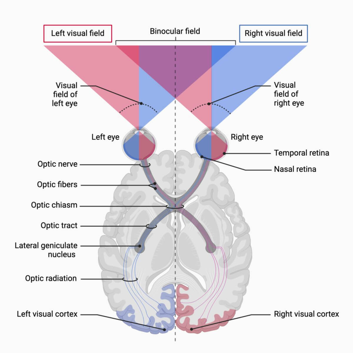
The optic chiasm is a partial crossing point for the nerve fibers of the optic nerve.
- The nerve fibers from the nasal retinal section of the right eye, which represent the temporal right visual field, cross over to the left side in the optic chiasm.
- The nerve fibers from the temporal retinal section of the left eye, which represent the nasal left visual field, stay on the left side.
Conversely, the same pattern occurs on the other side:
- The nerve fibers from the nasal retinal section of the left eye, which represent the temporal left visual field, cross over to the right side in the optic chiasm.
- The nerve fibers from the temporal retinal section of the right eye, which represent the nasal right visual field, stay on the right side.
3. Physiology
The optic chiasm is a critical part of the visual pathway. Due to the partial crossing of optic nerve fibers, the right cerebral hemisphere processes visual information from the left half of the visual field, while the left hemisphere processes visual information from the right half of the visual field.
4. Clinic
Lesions of the optic chiasm can cause bitemporal hemianopsia which is characterized by the loss of both temporal visual fields. This occurs because the nasal fibers of the optic nerve, which represent the temporal visual fields, cross at the optic chiasm.