Corpus: Eye


1. Definition

2. Background
The simplest "eyes" are light-sensitive sensory cells on the outer skin that function as passive optical systems, merely detecting light or darkness. This is known as the skin's sense of light.
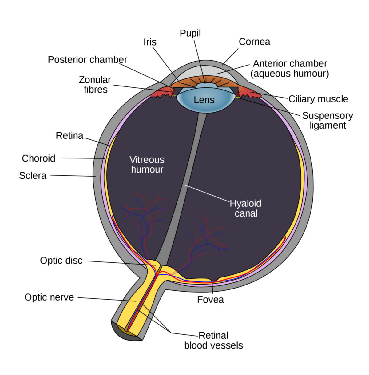
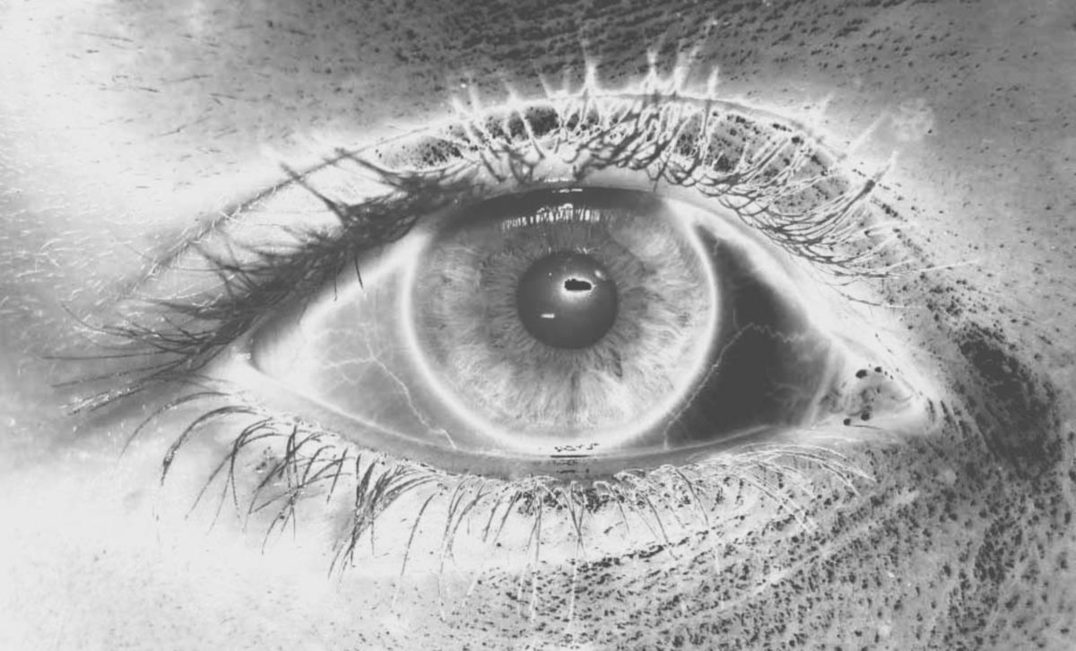
In most vertebrates and some mollusks, such as squid, an image is projected onto the retina, a light-sensitive layer in the eye. Sensory cells in the retina perceive the light, and nerve impulses are transmitted to the brain via the optic nerve. Generally spherical, the eye contains a gel-like, transparent substance called the vitreous body. It typically has a variable lens for image focusing and an iris, a ring muscle that can shrink the pupil to protect the eye from excessive light. The eye is often protected by eyelids and located deep in the skull with bony ridges for added protection.
3. Evolution
Although the eyes of vertebrates and mollusks are structurally similar, they have evolved independently. In vertebrates, the eye develops from an evagination of brain-forming cells, while in mollusks, it forms from an invagination of the outer cell layer that becomes the skin. Eyes of various designs have evolved approximately 40 times throughout evolution.
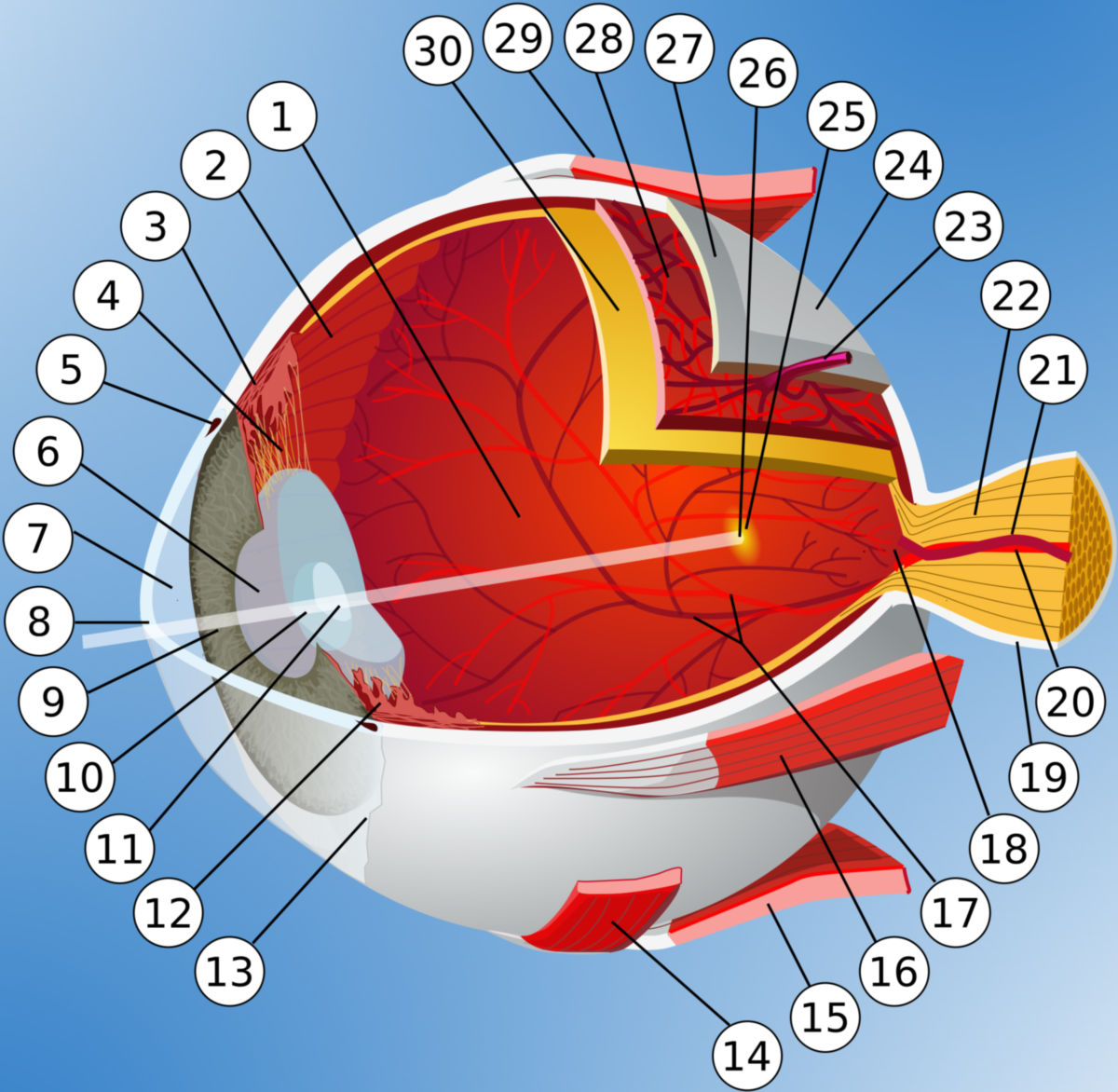
4. Anatomy
The eye can be divided into the eyeball and its appendages (eyelids, eye muscles, etc.), located in the eye socket (orbit). The ocular bulb consists of three layers:
- Fibrous layer of the eyeball (outermost layer)
- Sclera
- Cornea
- Uvea (middle layer)
- Iris
- Ciliary body
- Choroid
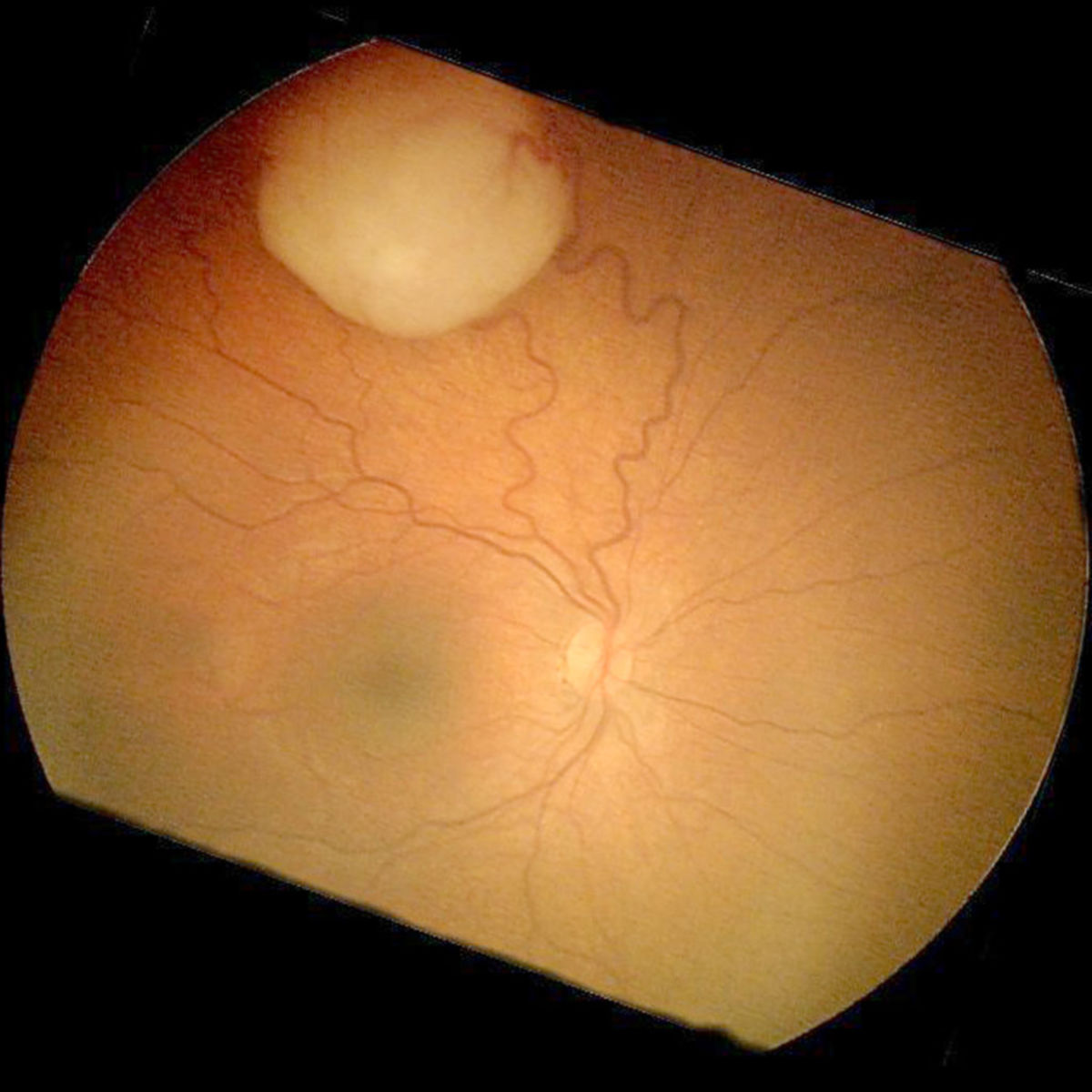
- Retina (innermost layer)
The interior of the eyeball contains the vitreous body and the crystalline lens as light-refracting structures. In the anterior section of the eye, a distinction is made between the two eye chambers (anterior and posterior chambers).
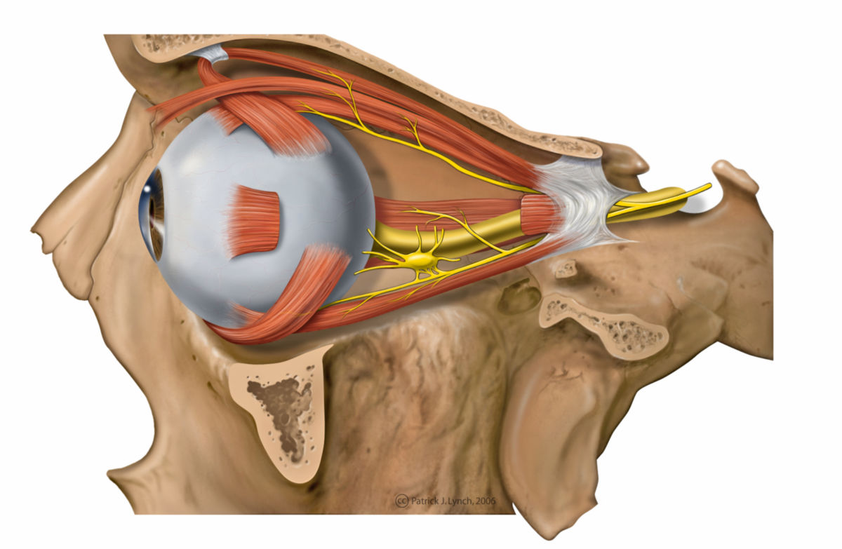
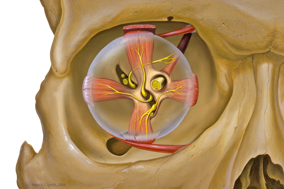
4.1. Eye musculature
The eye musculature can be categorized into the outer and the inner eye musculature.
The outer eye muscles, which are skeletal muscles under voluntary control, are responsible for changing the direction of gaze. In contrast, the inner eye muscles are smooth muscles that function in the context of accommodation and pupillomotor function.
In a broader sense, mimic muscles located around the outer eye can also be considered part of the eye musculature, as they regulate the width of the eyelids.
4.2. Vascular supply
The arterial supply of the eye comes from branches of the ophthalmic artery:
- Central retinal artery
- Posterior ciliary arteries
- Anterior ciliary arteryThe central retinal artery supplies the optic nerve and the inner layers of the retina, while the ciliary arteries supply the choroid and, through it, the outer layers of the retina. The blood-retina barrier exists between the choroid and the retina.
Venous drainage is primarily handled by the following vessels, all of which drain into the superior and inferior ophthalmic veins:
4.3. Innervation
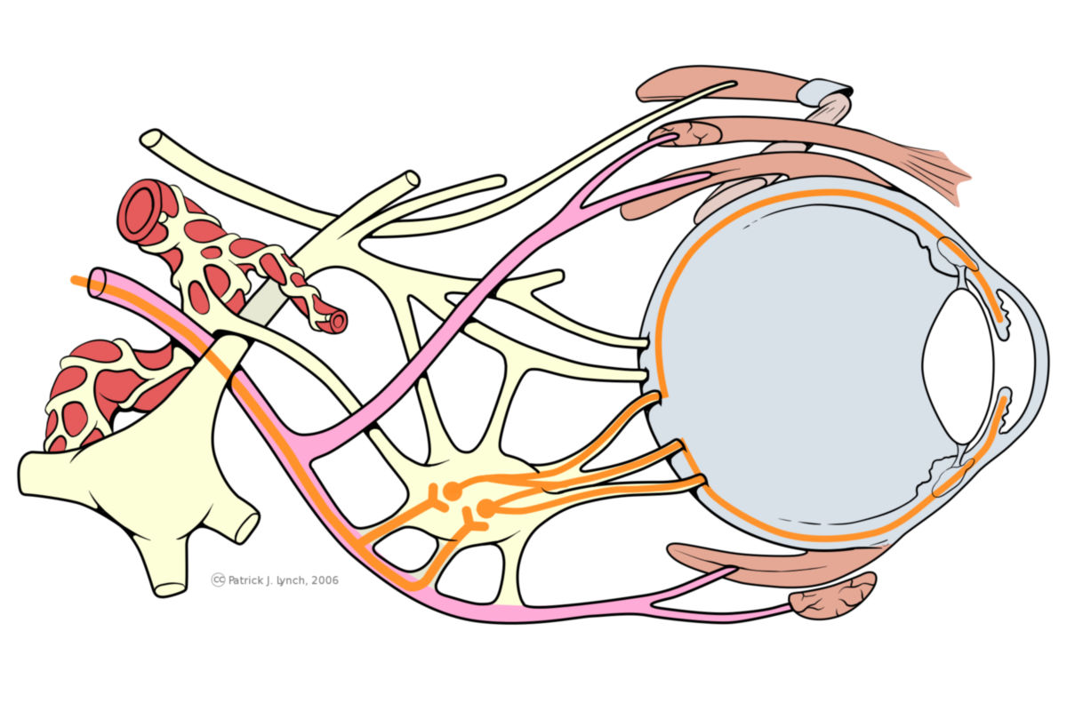
The eye is primarily innervated by various cranial nerves:
- Optic nerve: It is the first section of the visual pathway, transmitting visual information from the eye to higher brain centers.
- Oculomotor nerve: Controls four external eye muscles (superior rectus, medial rectus, inferior rectus, and inferior oblique) and the superior levator palpebrae muscle. It also carries visceromotor fibers to the ciliary ganglion.
- Trochlear nerve: Innervates the superior oblique muscle with somatomotor fibers.
- Abducens nerve: Supplies the lateral rectus muscle with somatomotor fibers.
- Long ciliary nerves: These sympathetic fibers from the superior cervical ganglion innervate the iris dilator muscle, which dilates the pupil.
- Short ciliary nerves: These carry parasympathetic fibers from the oculomotor nerve, which are switched in the ciliary ganglion. The short ciliary nerves innervate the iris sphincter muscle (for pupil constriction) and the ciliary muscle (for accommodation).
5. Physiology
5.1. Visual acuity
Visual acuity is the ability of an organism to perceive and recognize various environmental structures through the eye. The higher the visual acuity, the smaller the distance at which two points can be perceived as separate. Visual acuity is a measurable variable used as a diagnostic parameter in eye examinations.
5.1.1. Accommodation
Accommodation is the process of focusing on objects at different distances. Focusing on distant objects is referred to as distance accommodation, while focusing on near objects is referred to as near accommodation. The ciliary muscle is responsible for this process. Its contraction leads to the slackening of the zonular fibers, which anchor the lens to the ciliary body and keep it under tension. When the tension of the zonular fibers is reduced, the lens follows its own elasticity, becomes rounded, and changes its refractive power.
5.1.2. Field of Vision
The field of vision is the area that can be seen when the head is held still and the gaze is directed straight ahead without movement. In humans, the binocular horizontal field of vision is approximately 210°, expressed in degrees of visual angle.
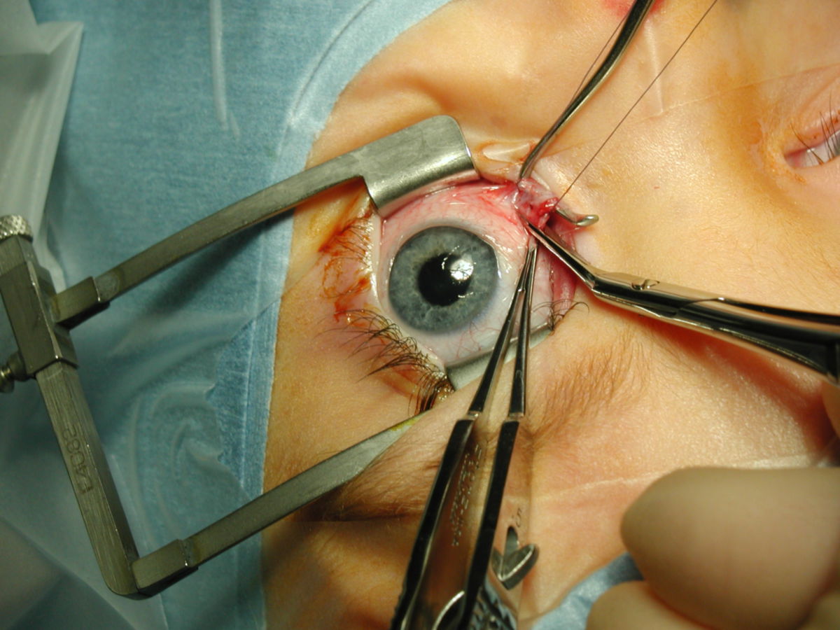
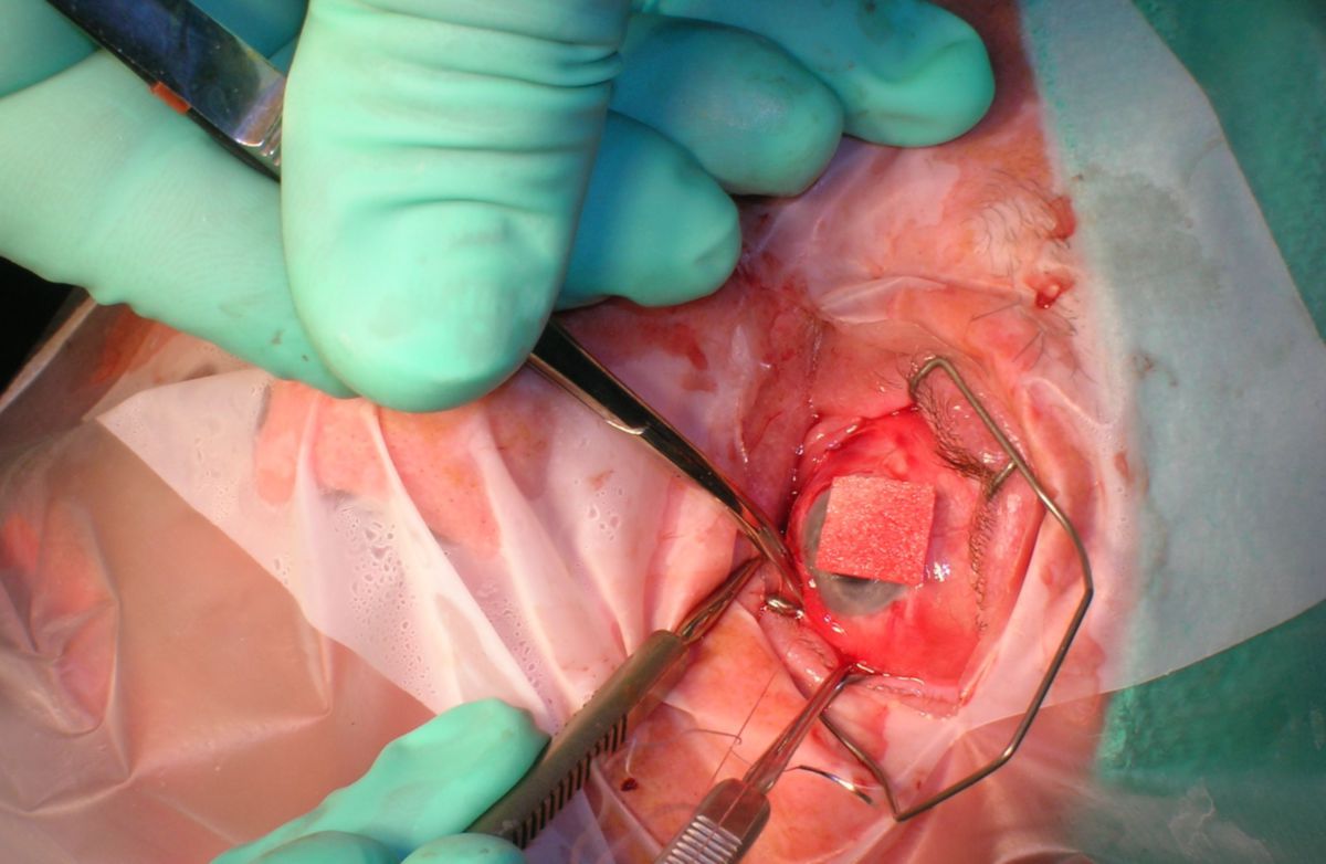
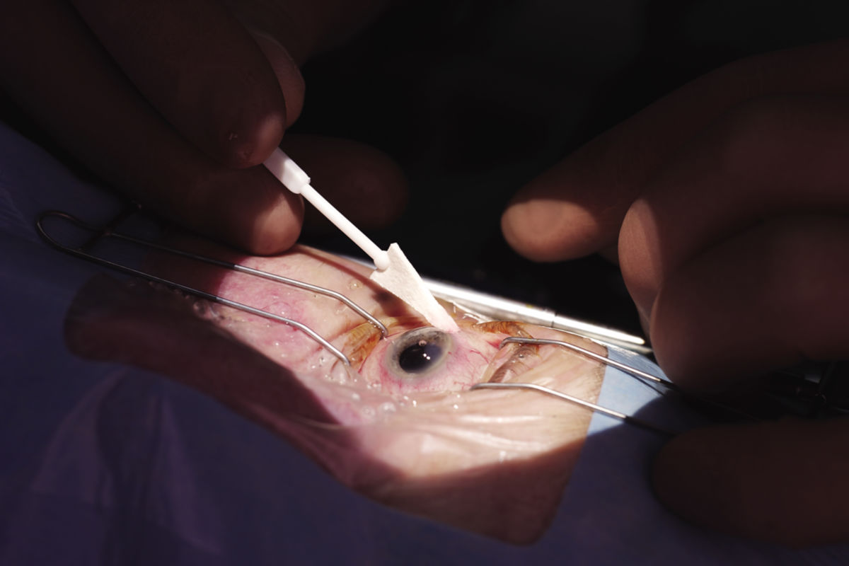
6. Clinic
Diseases of the eye fall under the domain of ophthalmology. In university ophthalmology, a distinction is made between diseases of the anterior and posterior segments of the eye. Common and significant eye diseases include:
- Corneal curvature (astigmatism)
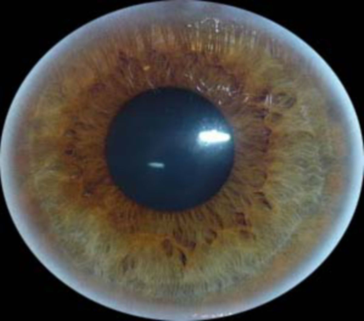
- Color blindness
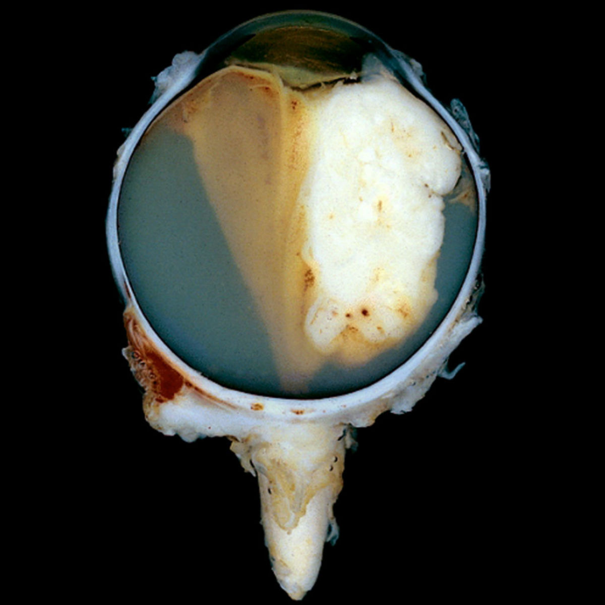
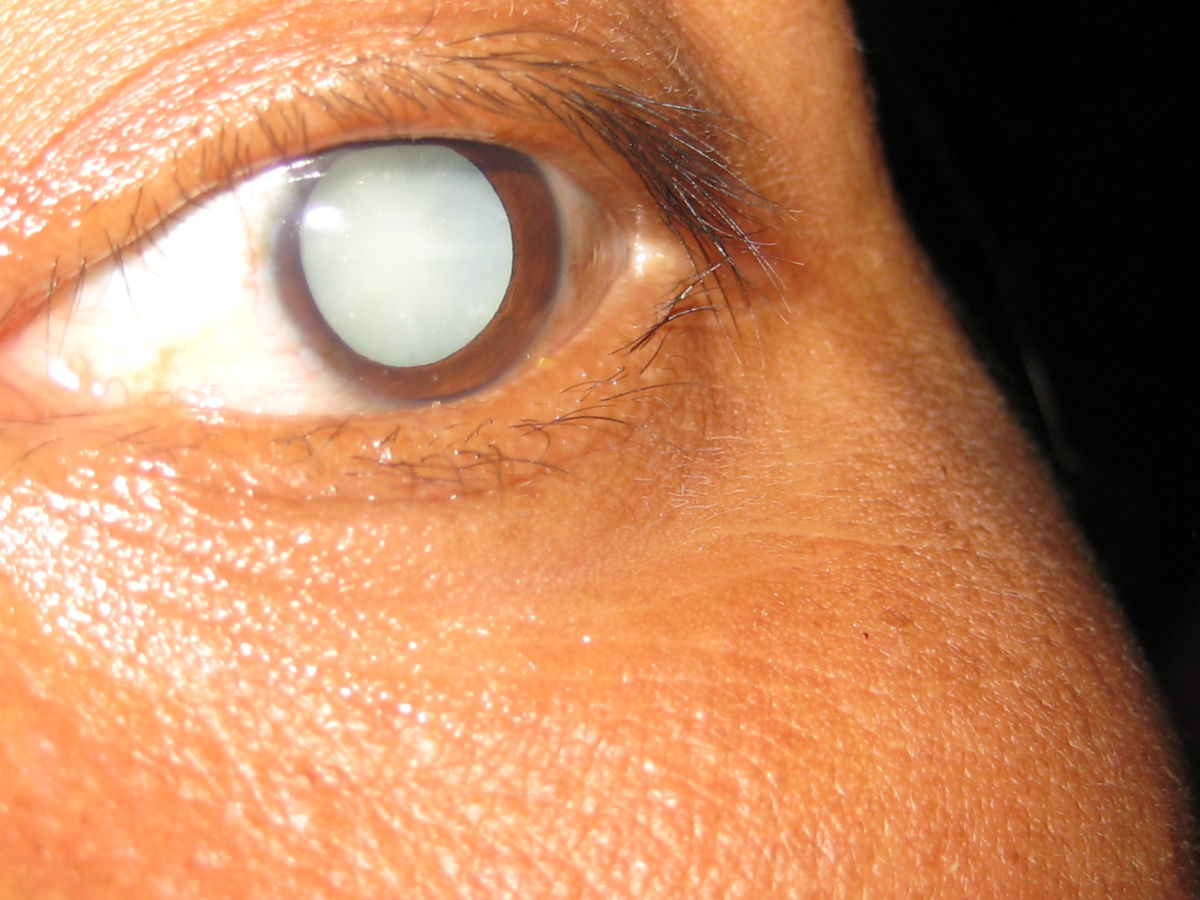
- Cataracts (grey cataract)
- Glaucoma
- Nearsightedness (myopia)
- Farsightedness (hyperopia)
- Age-related farsightedness (presbyopia)
- Loss of a field of vision (scotoma)
- Misalignment of the eyes (strabismus)
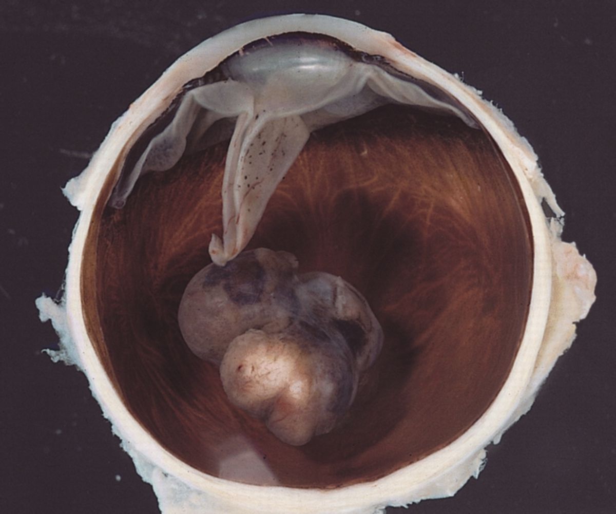
Less common but clinically significant are tumor diseases of the eye.