Corpus: Brain


1. Definition
The brain is part of the central nervous system (CNS) in vertebrates and is located in the head, within the cranial cavity. It serves as the central control center of the body.
2. Evolution
The vertebrate brain is responsible for processing complex sensory information and coordinating intricate behaviors. It serves as the primary site for integrating vital information within an organism. Together with the spinal cord, the brain forms the central nervous system.
In addition to vertebrates, some invertebrates, like octopuses, also possess highly complex brains that allow them to perform specialized tasks. In broader terms, the central nervous system in various invertebrates, such as annelids or insects, is sometimes referred to as a brain. Depending on the structure, these are called the cerebral ganglion, upper cervical ganglion, or similar terms.
The human brain is the most extensively studied brain in the animal kingdom, along with simpler nervous systems found in some worms.
3. Embryology
3.1. General principles
The development of the brain begins with the closure of the neural tube. At the cranial end of the neural tube, three brain vesicles form.
The wall of the neural tube consists of undifferentiated neural epithelial cells. These cells divide and give rise to two new cell types: primitive nerve cells (neuroblasts) and primitive support cells (glioblasts).
Neuroblasts develop into motor neurons of the anterior (ventral) horn and sensory neurons of the posterior (dorsal) horn.
The neuroblasts migrate into the mantle layer, leading to the formation of two thickenings: the motor plate (ventral thickening) and the sensory plate (dorsal thickening). Between these, a groove called the sulcus limitans forms, which later contributes to the neurons of the autonomic nervous system.
The roof plate lies between the two sensory plates, while the floor plate is located between the two motor plates. Surrounding the sensory and motor plates is the marginal zone.
After neuroblast formation is complete, glioblasts develop and migrate into the mantle layer. They differentiate into two types of supporting cells: protoplasmic astrocytes and fibrillar astrocytes.
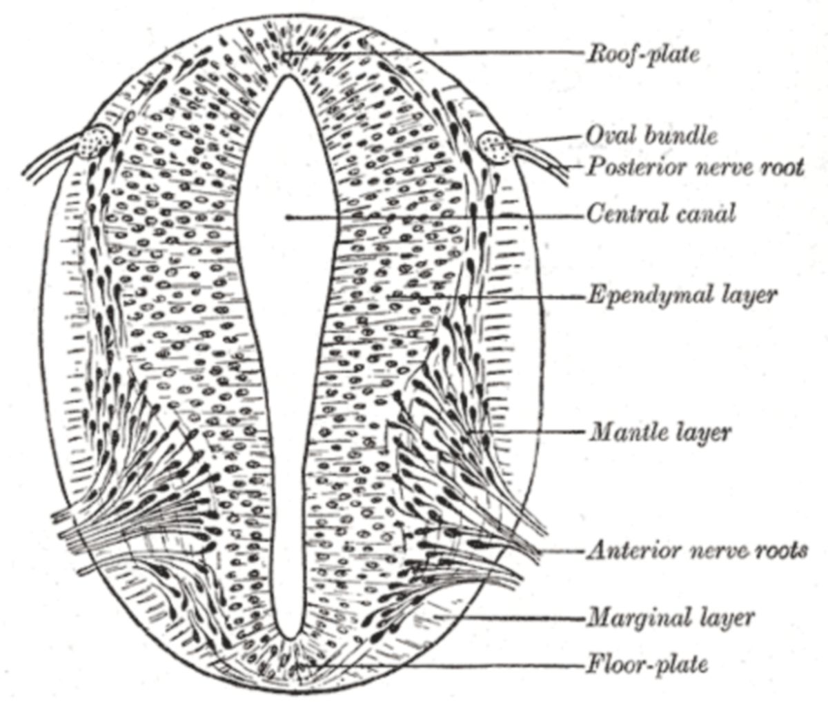
Once the neural epithelial cells have stopped forming neuroblasts and glioblasts, they develop into ependymal cells.
3.2. Special embryology
The neural tube consists of two main parts: the cranial portion, which contains the cerebral vesicles, and the caudal portion.
The three primary regions of the brain develop from the cerebral vesicles: the forebrain (prosencephalon), midbrain (mesencephalon), and hindbrain (rhombencephalon). The spinal cord develops from the caudal part of the neural tube.
The development of these brain regions is guided by five key structures of the neural tube: the base plate, wing plate, roof plate (cover plate), floor plate, and marginal zone. Each of these structures differentiates uniquely in different segments of the neural tube.
In the caudal part of the neural tube (which gives rise to the spinal cord), the ependyma forms a lining around the central canal. In the cranial part of the neural tube (the region of the cerebral vesicles), the central canal transforms into the ventricular system. This ventricular system is lined by ependymal cells, which are responsible for producing cerebrospinal fluid.
3.2.1. Forebrain (Prosencephalon)
The forebrain gives rise to the diencephalon and the telencephalon. These structures form through complex differentiation processes and the rotation of the brain hemispheres. During development, the hemispheric vesicles (one on each side of the brain) expand unevenly, predominantly in a caudal and basal direction. This uneven growth leads to the formation of the temporal lobe. The hemispheres also shift through a forward-upward and backward-downward movement.
3.2.2. Midbrain (Mesencephalon)
The midbrain develops into the structures known as the crura cerebri, the tegmentum, and the tectum.
3.2.3. Hindbrain (Rhombencephalon)
The hindbrain develops into two key redions: the metencephalon, which gives rise to the cerebellum and pons and the myelencephalon, which forms the medulla oblongata.
4. Anatomy
4.1. Macroanatomy
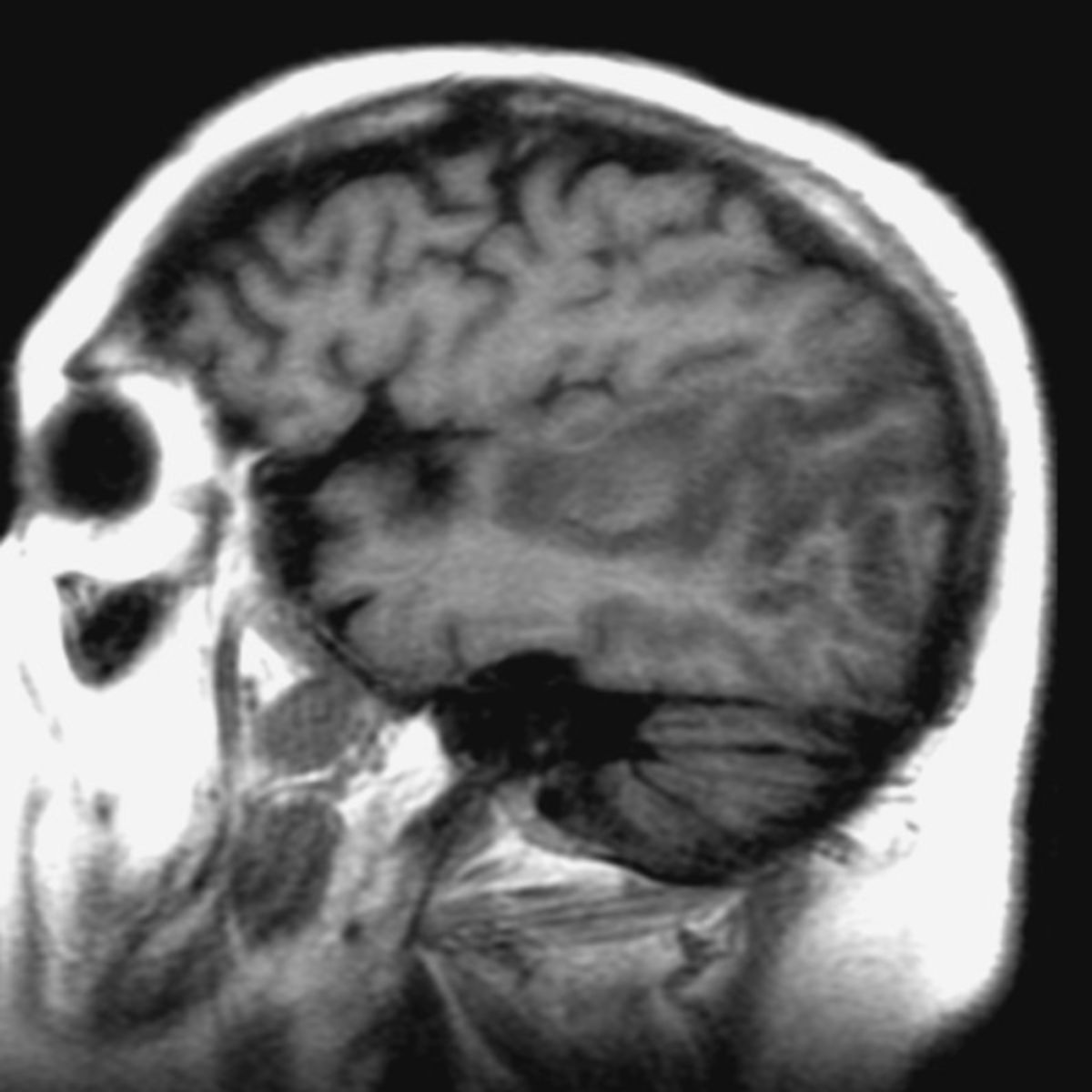
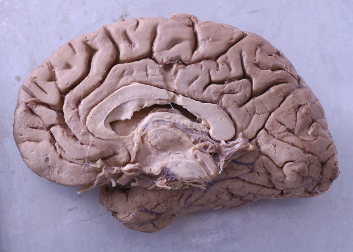
The human brain has an approximately oval shape when viewed from above, and its tissue appears greyish-yellow. Externally, the undivided brain can be separated into three main sections: the prominently bulging cerebrum, the cerebellum, and the hindbrain, which transitions into the spinal cord.
The cerebrum is divided into two equal halves, called hemispheres, by a central groove known as the longitudinal fissure. The surface of the cerebrum is characterized by cerebral convolutions (gyri) and intervening grooves (sulci), which together form a distinct pattern. Within the gyri and sulci lie various functional centers of the brain, such as the auditory cortex in the temporal lobe and the visual cortex in the occipital lobe.
The average weight of the brain ranges between 1,300 and 1,600 grams.
4.1.1. Brain parts
- Prosencephalon (forebrain)
- Telencephalon (end brain)
- Cortex
- Basal ganglia
- Limbic system
- Diencephalon
- Thalamus
- Epithalamus
- Subthalamus
- Hypothalamus
- Metathalamus
- Telencephalon (end brain)
- Mesencephalon (midbrain)
- Tectum
- Segmentum
- Crura cerebri
- Rhombencephalon (rhomboid brain)
- Metencephalon (hindbrain)
- Cerebellum (cerebellum)
- Pons
- Myelencephalon (hindbrain)
- Medulla oblongata
- Metencephalon (hindbrain)
- (spinal cord)
The medulla oblongata, pons and mesencephalon are summarised as the brain stem (encephalic trunk).
4.2. Microanatomy
Each section of the brain, derived from its respective cerebral vesicle, develops its own unique structure.
However, since the cerebral cortex constitutes the largest portion of the brain by volume, two key structural classifications are highlighted here: the isocortex and the allocortex.
These classifications do not represent separate areas of the brain, but rather describe different structural types within the cerebral cortex.
5. Physiology
5.1. Cerebral blood flow
Cerebral blood flow is essential for supplying oxygen and nutrients to the brain's nerve cells. In healthy adults, about 15 % of the total cardiac output is directed to the brain and its surrounding tissue, which corresponds to approximately 700 ml of blood per minute.
5.2. Cerebral metabolism
Under normal conditions, the brain primarily relies on glucose and oxygen to meet its metabolic needs. However, during periods of high plasma concentrations of ketone bodies (e.g., fasting or ketoacidosis), the brain can utilize these molecules for energy production. Despite this adaptation, ketone bodies can only provide up to approximately 50 % of the brain's energy requirements.
5.3. Brain functions
Not all sensory information reaches the cerebral cortex and becomes part of conscious awareness. Many signals are pre-processed unconsciously by peripheral nerve plexuses or centers in the brainstem. Reflex arcs handle tasks that require rapid responses and do not involve conscious registration.
Humans also possess an autonomic nervous system, which regulates vital involuntary functions such as breathing, circulation, food intake, digestion and excretion, fluid balance, and reproduction. Conscious control of all these processes would overwhelm the brain structures responsible for perception and decision-making, effectively rendering them nonfunctional.
The structure—and, to a lesser extent, the size—of the brain can serve as an indicator of an animal's capacity for learning and intelligence. However, the ability to learn is not solely reliant on the brain itself. Neuronal plasticity, or the ability of the nervous system to adapt and change, occurs at nearly all hierarchical levels of the nervous system.
6. History of brain research
- Egypt: "Textbook of Surgery" with description of brain furrowing, recognition of brain injuries (deviating eye position, dragging of a foot, loss of speech)
- Hippocrates: epilepsy can be triggered by stimuli
- Galen: first neurophysiological experiments (incisions, lesions)
- Andreas Vesalius: brain anatomy
- Descartes: dichotomy of body and soul
- Thomas Willis: grey/white matter
- Gall: phrenology (skull mapping)
- Paul Broca: localised motor speech centre in the left frontal lobe
- Wernicke: localised sensory speech centre in the left temporal lobe
- Brodmann: categorisation of the cerebral cortex into 52 areas
- Santiago Felipe Ramón y Cajal: neuron theory