Corpus: Maxilla


Synonym: upper jaw
1. Definition
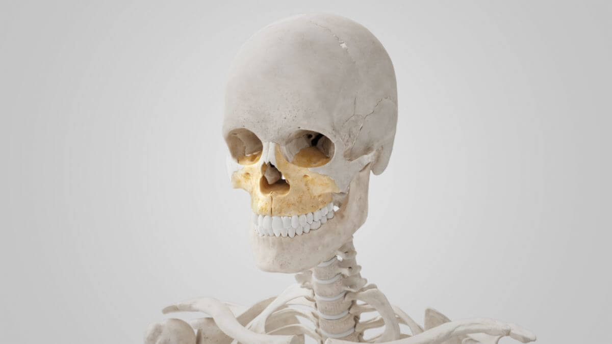
The maxilla is the largest bone of the facial skull after the mandible. The right and left maxilla together form the upper jaw.
The bone forms the boundary walls of three important body cavities: the roof of the oral cavity, the floor and side wall of the nasal cavity, and the floor of the orbit.
2. Anatomy
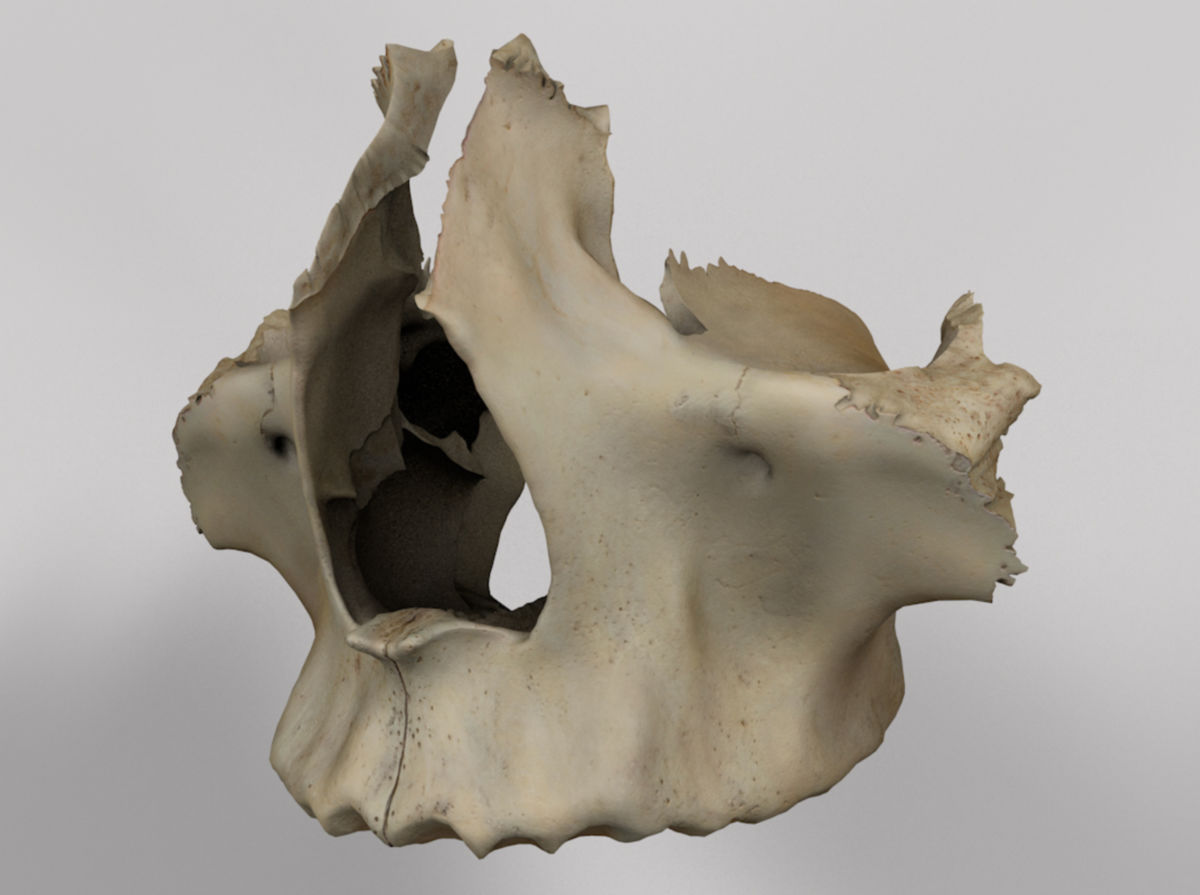
The maxilla can be anatomically divided into a body (corpus maxillae) and its bony processes: the zygomatic process, the frontal process, the alveolar process, and the palatine process.
2.1. Body of the maxilla
2.1.1. Anterior surface
The anterior surface of the maxilla faces forward and laterally. It features a series of bony elevations that mark the position of the tooth roots of the maxilla. Above the roots of the incisors is a depression called the incisive fossa, which serves as the origin of the depressor septi nasi muscle and the alar part of the nasalis muscle. Caudal to the incisive fossa is the origin area of the orbicularis oris muscle.
Adjacent to the incisive fossa is a larger depression called the canine fossa, separated by a vertical bone ridge known as the canine eminence, which corresponds to the long root of the canine tooth. The levator anguli oris muscle originates from this area. Above the canine fossa lies the infraorbital foramen, which transmits the infraorbital nerve and vessels. The lower orbital rim, located above the foramen, serves as the origin of the levator labii superioris muscle. Medially, the anterior surface of the maxilla ends at the piriform aperture, with the bony edge forming the anterior nasal spine.
2.1.2. Intratemporal surface
The infratemporal surface forms part of the infratemporal fossa. It is convex, facing laterally and posteriorly. It is separated from the anterior surface by the zygomatic process and a bony ridge running upwards from the first molar. This surface has openings for the alveolar canals, which carry branches of the superior posterior alveolar artery and nerve. The caudal section features the maxillary tuberosity, roughened on its lateral side for articulation with the pyramidal process of the palatine bone and serving as an origin for fibers of the medial pterygoid muscle. Above the maxillary tuberosity is a smooth surface forming the anterior border of the pterygopalatine fossa, containing a cavity for the maxillary nerve.
2.1.3. Orbital surface
The orbital surface is triangular and smooth, forming the largest part of the orbital floor. It is bordered medially by an irregular edge with a notch at the front called the lacrimal notch. The maxilla articulates with the lacrimal bone, the orbital plate of the ethmoid bone, and the orbital process of the palatine bone. Posteriorly, the orbital surface is bordered by a rounded edge forming the anterior edge of the inferior orbital fissure. The infraorbital groove, a prominent structure, runs along this surface, carrying the infraorbital artery, vein, and nerve. The groove leads to the infraorbital canal, ending at the infraorbital foramen on the bone surface.
2.1.4. Nasal surface
The nasal surface leads into the maxillary sinus with an irregular bone edge. The concave surface below this opening forms part of the inferior meatus of the nasal cavity. Behind this, a rough area articulates with the perpendicular plate of the palatine bone. The caudal part of this area features a small trench forming the pterygopalatine canal when covered by the palatine bone. The nasal surface also has a flat section forming part of the middle nasal meatus, separated by a small oblique bone ridge called the conchal crest, which articulates with the inferior nasal concha. The lacrimal groove can be seen at the upper end of the conchal crest, forming the nasolacrimal canal when closed by the lacrimal bone and inferior nasal concha.
2.2. Zygomatic process
The zygomatic process is a strong, triangular bony projection positioned at an angle between the anterior, orbital, and infratemporal surfaces of the maxilla. It ends in a rough, serrated surface articulating with the zygomatic bone and forms part of the infratemporal fossa dorsally. The zygomaticoalveolar crest is a bony ridge at its base.
2.3. Frontal process
The frontal process is a bony plate that extends upwards, medially, and posteriorly next to the nose. Its lateral side is smooth, merging with the anterior surface of the maxilla, and serves as the origin of the levator labii superioris muscle, the orbicularis oculi muscle, and the medial palpebral ligament. The medial surface forms part of the lateral nasal wall, articulating with the ethmoid bone and forming the crista ethmoidalis. The superior edge articulates with the frontal bone, the anterior edge with the nasal bone, and the posterior margin with the lacrimal groove.
2.4. Alveolar process
The alveolar process is a thick, horseshoe-shaped bony ridge surrounding the bony palate, containing the dental compartments for the teeth. The size and shape of these compartments vary with the type of tooth enclosed. The canine tooth compartment is the deepest. The roots of the incisors and canines create vertical elevations on the facial surface called alveolar juga. The buccinator muscle originates behind the first molar on this surface.
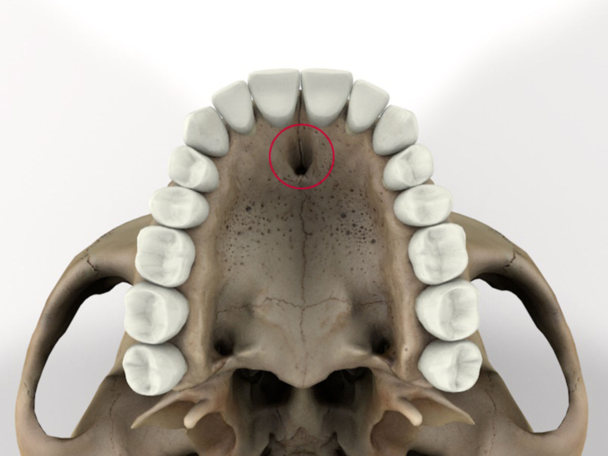
2.5. Palatine process
The palatine process is a prominent bony projection forming a large part of the nasal floor and hard palate. Its inferior surface is rough and concave, perforated by numerous foramina for the passage of blood vessels and nerves. The incisive foramen is the most notable structure, located directly behind the incisors, with canals for the descending palatine artery and nasopalatine nerve. The median palatine suture marks the junction of the two palatine processes, while the transverse palatine suture delineates the border with the palatine bone.
The superior surface of the palatine process is smooth and concave, contributing significantly to the bony floor of the nose and forming the nasal crest with its counterpart. The anterior nasal spine is a pointed projection at the rostral end, and the lateral edge merges into the maxilla.