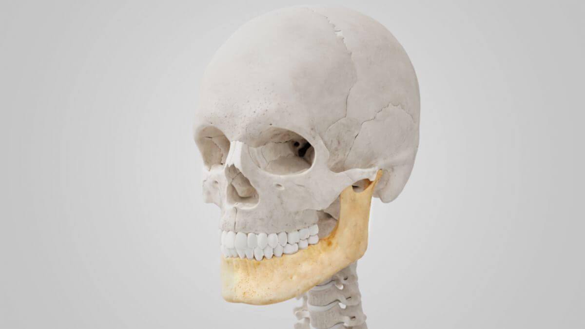
Corpus: Mandible


Synonym: lower jaw
1. Definition
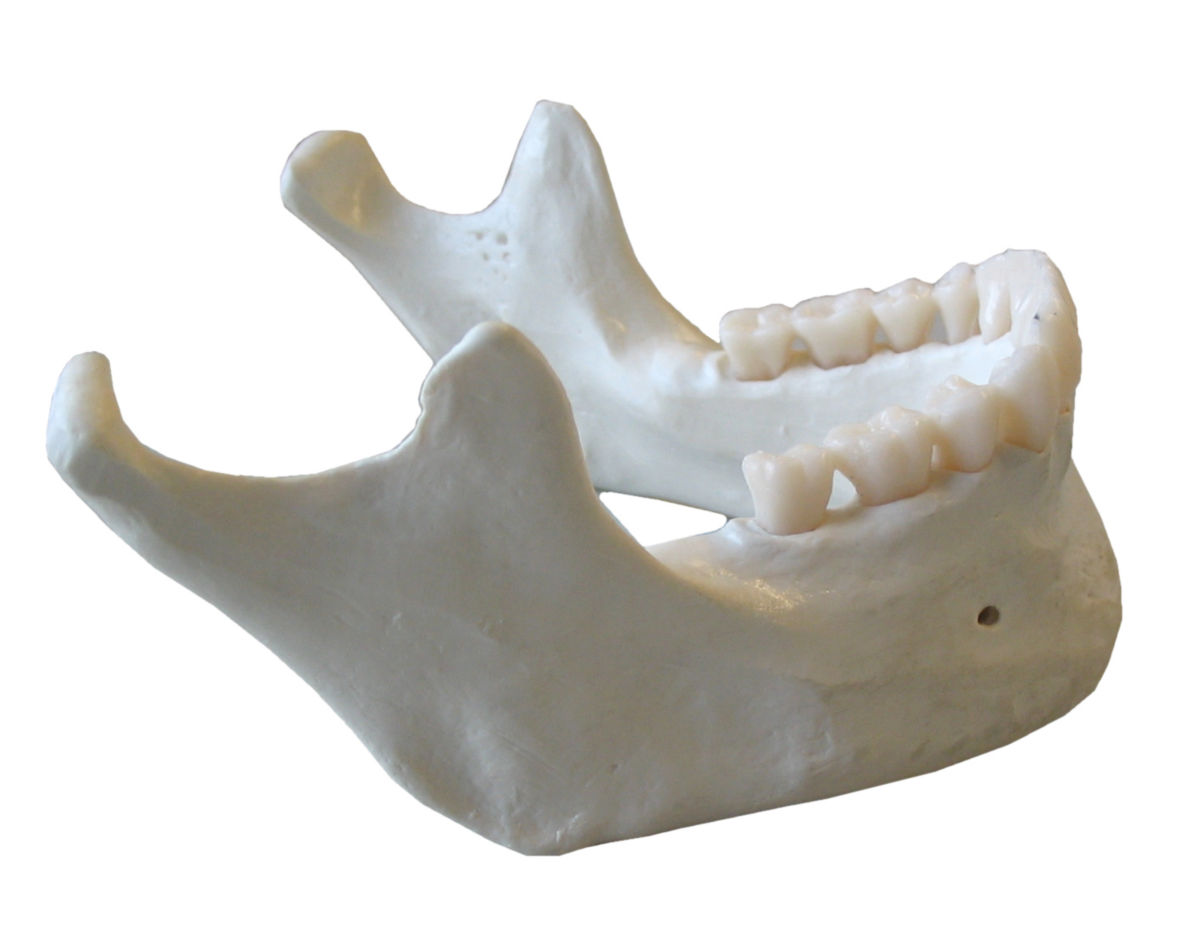
2. Body of the mandible
The body of the mandible has a horseshoe shape. Its upper boundary is the alveolar process, which contains the 16 bony tooth compartments and can vary considerably in shape depending on the dentition. The alveolar process almost completely disappears with tooth loss in old age, representing a physiological change rather than a variety.
The lower edge of the body of the mandible merges dorsally into the angle of the mandible on both sides. The body has two surfaces:
2.1. External surface
The external surface is the convex side of the mandible. In the median line, a small bone ridge marks where the right and left parts of the lower jaw bone fuse during the embryonic period, called the symphysis. The bony ridge divides caudally along its course and merges into a triangular elevation, the mental protuberance. This elevation has a prominence on the left and right (mental tubercle), which merges into a fine ridge running diagonally across the bone (linea obliqua).
The linea obliqua runs cranially and dorsally to the anterior edge of the respective mandibular ramus. The triangularis muscle and the depressor labii inferioris muscle originate from it, and the platysma attaches caudally to it. Cranial to the linea obliqua on the alveolar process, directly below the molars, is the insertion of the buccinator muscle.
Below the incisors is the incisive fossa, which serves as the origin of the mentalis muscle and part of the orbicularis oris muscle. Caudal to the second premolar, the mental nerve, artery, and vein emerge on both sides in the mental foramen.
2.2. Internal surface
The concave side of the mandible is the internal surface. Near the lower part of the symphysis, small protrusions, the mental spines, represent the origin of the genioglossus and geniohyoid muscles. These spines can also form a bony ridge or a tuberosity. Below the spine lies the small oval digastric fossa, the origin of the digastric muscle. The mylohyoid line rises dorsally and cranially on both sides, mimicking the course of the external linea obliqua. It is the origin of the mylohyoid muscle, and in the posterior part, of the superior pharyngeal constrictor muscle. Above and below the linea, two depressions accommodate the sublingual gland and the submandibular gland.
3. Mandibular ramus
The mandibular ramus, which originates on both sides of the mandibular angles, has a square shape with two surfaces, four edges, and two projections. The entry point of the inferior alveolar nerve and the inferior alveolar artery into the alveolar canal, the mandibular foramen, is located on its inner side.
3.1. Coronoid process
The coronoid process is a thin, triangular bony projection flattened on both sides. Its anterior edge is convex and merges caudally into the anterior border of the mandibular ramus. The posterior margin is concave and forms the anterior margin of the mandibular notch. The lateral surface is smooth and serves as an attachment for the masseter muscle and the temporalis muscle. The medial surface also serves as the insertion for the temporalis muscle. It has a small bony ridge running anteroinferiorly to the last molar. Between this and the front edge of the mandibular ramus is a small triangular area, the retromolar triangle, serving as an attachment for the temporalis muscle and some fibers of the buccinator muscle.
3.2. Condylar process
The condylar process is more massive than the coronoid process and consists of two parts: the mandibular neck and the mandibular condyle.
Together with the intervening articular disc and the mandibular fossa of the temporal bone, the mandibular condyle forms the temporomandibular joint. It has a convex surface in both longitudinal and transverse directions. Its longitudinal axis points medially and slightly dorsally. A small tubercle on the lateral extension of the mandibular condyle serves as an attachment for the temporomandibular ligament.
The mandibular neck is relatively delicate and reinforced by bony ridges that descend laterally and frontally. The front is concave and forms the posterior border of the mandibular notch. The lateral pterygoid muscle attaches medially.
4. Biomechanics
Different movements of the mandible include:
- Protrusion: Advancement of the mandible from the resting position
- Retrusion: Retraction of the mandible from the resting position
- Protraction: Advancement of the mandible from the retrusion position to the resting position
- Retraction: Retraction of the mandible from the protrusion position to the resting position
- Laterotrusion: Lateral movement away from the midline
- Mediotrusion: Lateral movement towards the midline
Laterotrusion and mediotrusion refer to one half of the mandible and always occur together.