Corpus: Superior pharyngeal constrictor muscle


from Latin: constringere - to constrict and Greek: pharynx - throat
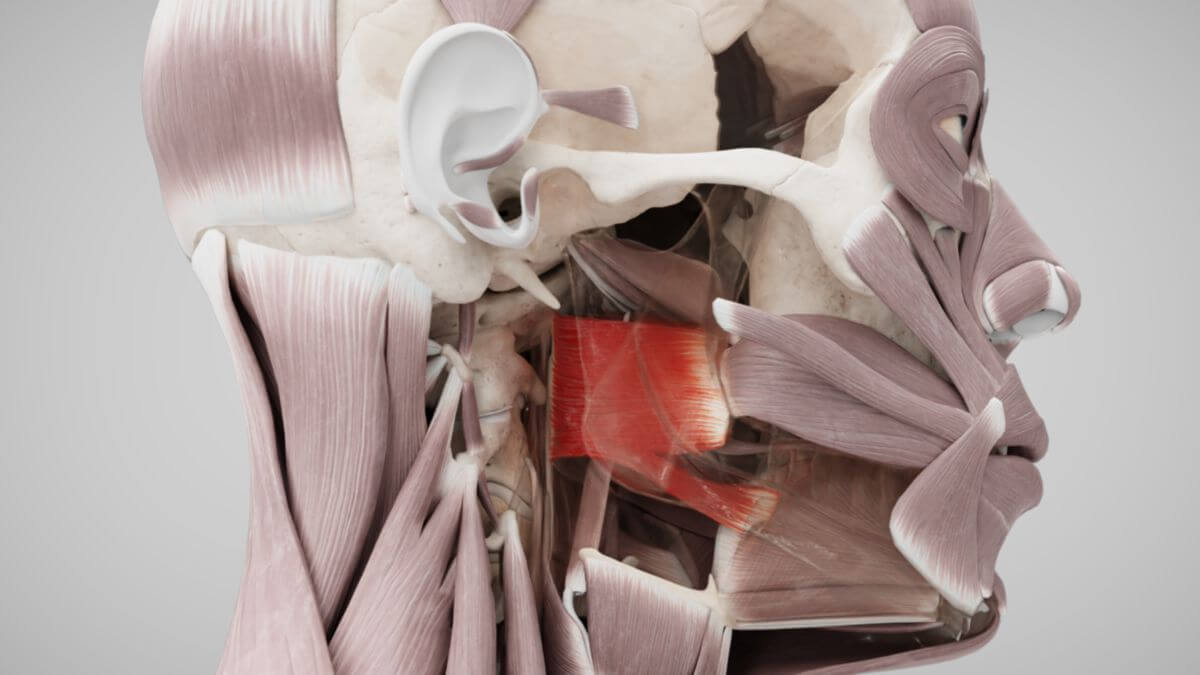
1. Definition
The superior pharyngeal constrictor muscle is a striated, branchiogenic muscle and the most cranial constrictor in the pharyngeal musculature. It is joined caudally by the middle pharyngeal constrictor muscle.
2. Embryology
The superior pharyngeal constrictor muscle predominantly develops from the muscle anlage of the third pharyngeal arch.
3. Anatomy
3.1. Classification
The superior pharyngeal constrictor muscle consists of four parts:
- Pterygopharyngeal part
- Buccopharyngeal part
- Mylopharyngeal part
- Glossopharyngeal part
3.2. Origin
The pterygopharyngeal part originates from the pterygoid hamulus of the sphenoid bone. The buccopharyngeal part originates from the pterygomandibular raphe. The mylopharyngeal part originates from the posterior part of the mylohyoid line of the mandible. The glossopharyngeal part originates from the muscular body of the tongue.
3.3. Insertion
The fibers of the superior pharyngeal constrictor muscle extend obliquely upward, medially, and backward, joining with the fibers of the muscle on the opposite side at the pharyngeal raphe. In the lower part, these fibers are overlapped by the upper fibers of the middle pharyngeal constrictor muscle in a tile-like manner. The upper fibers curve downward around the levator veli palatini muscle and the auditory tube.
4. Innervation
The superior pharyngeal constrictor muscle is innervated by the pharyngeal plexus, which consists of fibers from the glossopharyngeal nerve (cranial nerve IX) and the vagus nerve (cranial nerve X).
5. Function
The superior pharyngeal constrictor muscle constricts the nasopharynx during contraction, with the soft palate acting as an abutment. This action creates a thickening of the pharyngeal wall known as Passavant's ridge, which helps prevent food from entering the posterior sections of the nasal cavity during swallowing.