Corpus: Middle pharyngeal constrictor muscle


from Latin: constringere - to constrict and Greek: pharynx - throat
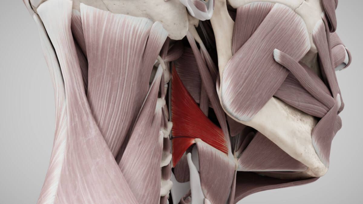
1. Definition
The middle pharyngeal constrictor muscle is a striated skeletal muscle, positioned as the middle of the three pharyngeal constrictors. It is joined cranially by the superior pharyngeal constrictor muscle and caudally by the inferior pharyngeal constrictor muscle.
2. Embryology
The middle pharyngeal constrictor muscle primarily develops from the muscle system of the 4th pharyngeal (gill) arch.
3. Anatomy
3.1. Classification
The middle pharyngeal constrictor muscle consists of two parts:
- the chondropharyngeal part
- the ceratopharyngeal part
3.2. Origin
The chondropharyngeal part originates from the lesser horn of the hyoid bone, while the ceratopharyngeal part originates from the greater horn of the hyoid bone.
3.3. Attachment
The fibers of the middle pharyngeal constrictor muscle extend obliquely upward, medially, and dorsally, merging with the fibers from the opposite side at the pharyngeal raphe. These fibers overlap the caudal fibers of the superior pharyngeal constrictor muscle in a manner similar to roof tiles.
4. Innervation
The middle pharyngeal constrictor muscle is innervated by the pharyngeal plexus, which includes contributions from the glossopharyngeal nerve (cranial nerve IX) and the vagus nerve (cranial nerve X).
5. Function
5.1. Swallowing
The middle pharyngeal constrictor muscle contracts to constrict the oropharynx (oral pharyngeal part), pushing the food bolus towards the esophagus during swallowing.
5.2. Phonetics
This muscle also plays a role in the formation of pharyngeal sounds, particularly in conjunction with the inferior pharyngeal constrictor muscle, and in the articulation of low, posterior vowels (such as "a").