Corpus: Hyoid bone


from ancient Greek: ῡ̔οειδής ("huoeides") - U-shaped
1. Definition
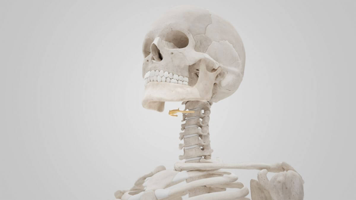
The hyoid bone is a small, horseshoe-shaped bone sometimes counted to the bones of the skull. It is connected via the right and left stylohyoid ligaments to the styloid process of the temporal bone and has no articular connection to other bones.
2. Anatomy
2.1. Overview
The hyoid bone is located approximately at the level of the third to fourth cervical vertebrae. The following relevant structures can be distinguished:
- Hyoid bone body (Corpus ossis hyoidei)
- Large hyoid horns (Cornua majora ossis hyoidei)
- Small hyoid horns (Cornua minora ossis hyoidei)
2.2. Musculature
The following muscles insert at the hyoid bone:
- Suprahyal muscles
- Infrahyoid muscles
In addition, the hyoid bone serves as the origin for
- Parts of the pharyngeal muscles
- Parts of the external muscles of the tongue
2.3. Ligaments
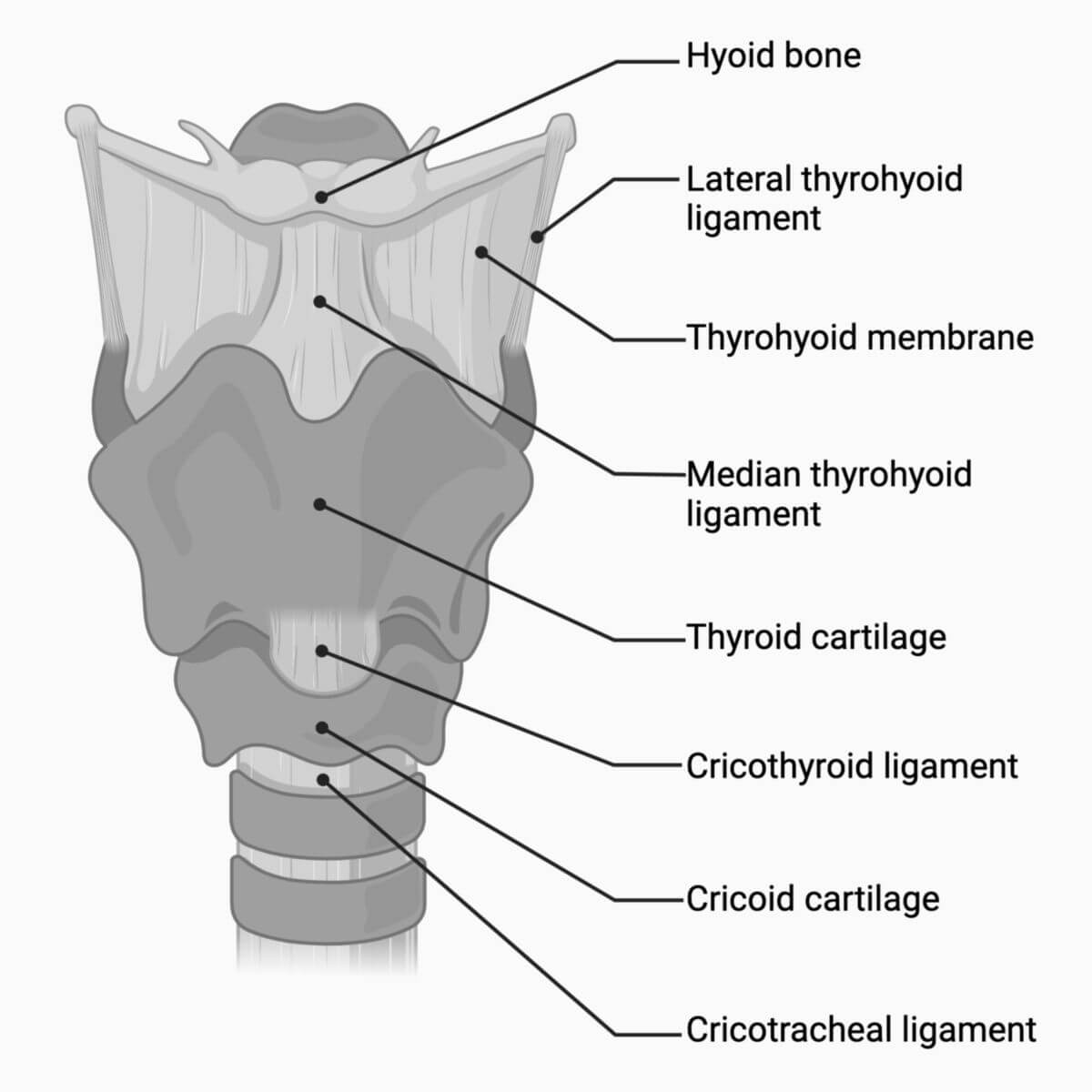
Numerous ligaments attach to the hyoid bone, transmitting movements to the larynx:
- Hyoepiglottic ligament: from the upper edge of the hyoid bone to the front of the epiglottis
- Median thyrohyoid ligament: from the body of the hyoid to the superior thyroid notch of the thyroid cartilage
- Lateral thyrohyoid ligament: runs from the lower edge of the large horns to the superior horns of the thyroid cartilage, containing the small triticeal cartilage
- Thyrohyoid membrane: fiber tracts between the lateral and median ligaments
The stylohyoid ligament attaches the hyoid bone cranially to the styloid process of the temporal bone.
2.4. Vascular supply
The hyoid bone is supplied with blood by the lingual artery.
3. Embryology
The small hyoid horns develop from the Reichert's cartilage of the second pharyngeal arch, while the large hyoid horns and the body of the hyoid bone develop from the third pharyngeal arch.
4. Ossification
Endochondral ossification of the hyoid bone begins at the small horns and occurs relatively late, involving a total of six ossification centers: one per horn and two in the hyoid body. After birth, the structure is still predominantly cartilaginous, with ossification completing by puberty at the latest.
5. Function
The hyoid bone transmits force between the supra- and infrahyoid muscles and the pharyngeal muscles during swallowing. It acts as a movable support and starting point for redirecting force. Due to its ligamentous attachment to the base of the skull, it stabilizes the larynx against gravitational force and muscle tension from the infrahyoid muscles.
6. Clinic
A hyoid fracture due to neck trauma is very rare due to the bone's flexibility. In forensic medicine, a hyoid fracture can indicate focused violence to the neck, such as in cases of hanging or strangulation.