Corpus: Thyroid cartilage


1. Definition
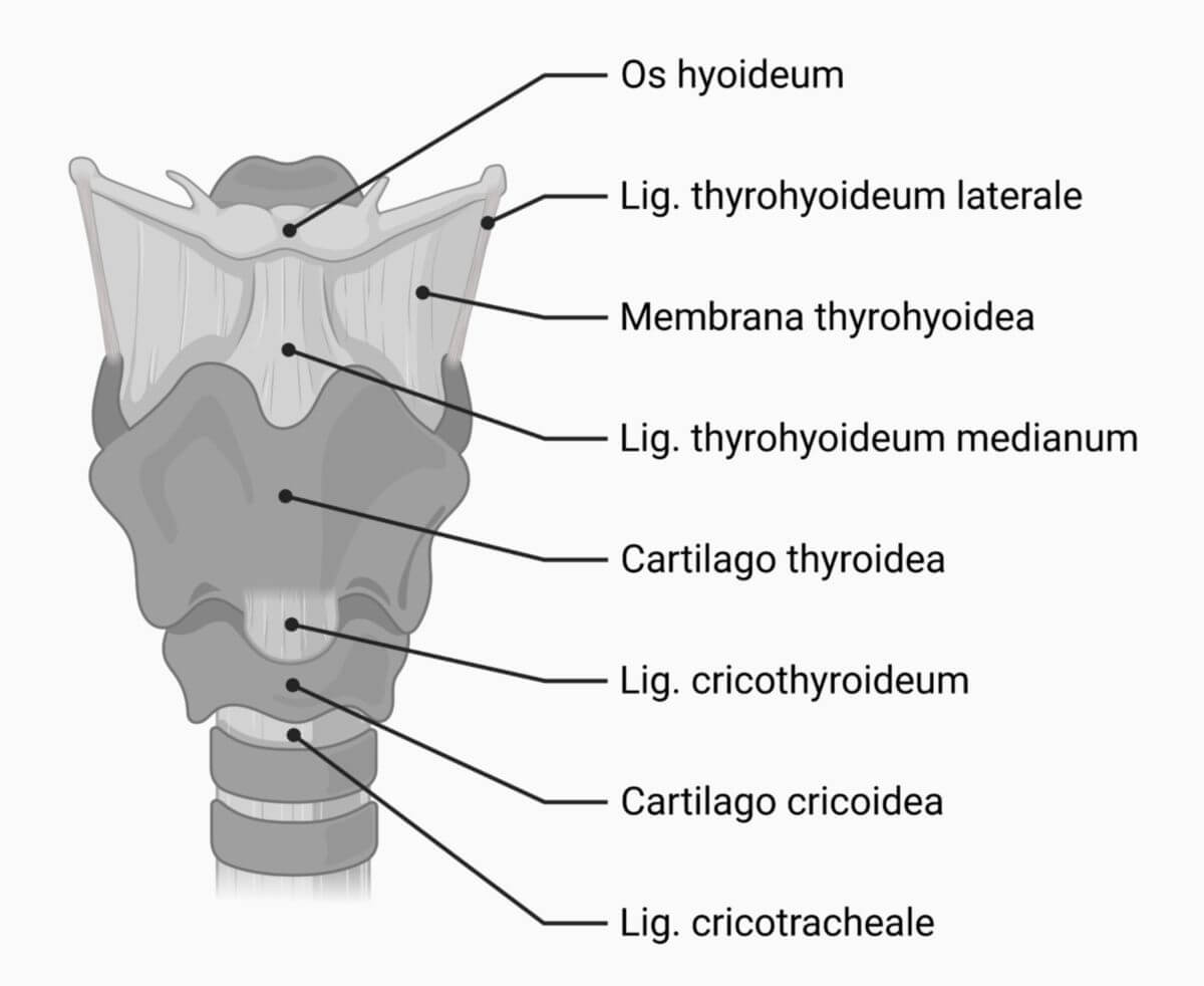
The thyroid cartilage is a cartilaginous part of the human larynx.
2. Anatomy
The thyroid cartilage consists of 2 cartilage plates (laminae) that are connected anteriorly in the center:
- Right lamina of the thyroid cartilage (Lamina dextra cartilaginis thyroideae)
- Left lamina of the thyroid cartilage (Lamina sinistra cartilaginis thyroideae)
Each lamina has a protrusion at its dorsal edge above and below, called the superior and inferior horn, respectively. Between the two laminae at the cranial end is a notch, the superior thyroid notch, which can be felt from the outside. At the end of the notch, the cartilage protrudes as the laryngeal prominence. Below the notch, the thyroepiglottic ligament connects the epiglottis stalk to the back of the thyroid cartilage.
At the caudal end of the thyroid cartilage is a less pronounced notch, the inferior thyroid notch. A diagonal line, the oblique line, runs along the two lateral surfaces of the thyroid cartilage, dividing the surfaces.
The thyroid cartilage forms the anatomical basis of the Adam's apple, which is more pronounced in men.
2.1. Muscle insertions
The following muscles originate or attach to the thyroid cartilage:
- Thyrohyoid muscle (in front of the oblique line)
- Sternothyroid muscle (behind the oblique line)
- Inferior pharyngeal constrictor muscle
- Cricothyroid muscleAdditionally, some fibers of the palatopharyngeal muscle insert at the posterior margin. On the inner surface:
3. Embryology
The thyroid cartilage develops, along with other laryngeal structures, from the anlagen of the fourth pharyngeal arch.
4. Histology
The cartilago thyroidea consists of hyaline cartilage. With age, it becomes increasingly calcified or ossified.
5. Clinic
In rare cases, a thyroid cartilage fracture can occur due to external force in the context of laryngeal trauma. The splitting of the thyroid cartilage during a surgical procedure is known as a thyroidectomy.