Corpus: Larynx


Synonym: voice box
1. Definition
The larynx is a structure made of cartilage, muscles, and connective tissues that functions as a valve, separating the windpipe (trachea) from the gullet (esophagus).
2. Anatomy
2.1. Topography
The larynx can be divided into three levels:
- The supraglottis (also known as the vestibule of the larynx) is the uppermost section. It extends from the entrance of the larynx (laryngeal inlet) to the gap between the vestibular folds (false vocal cords).
- The glottis (or the middle cavity of the larynx) is the middle section. It spans from the vestibular fold (rima vestibuli) to the true vocal cords (rima glottidis).
- The subglottis is the lowest section. It extends below the vocal cords and connects to the trachea at the lower border of the cricoid cartilage.
2.2. Cartilaginous framework
The shape of the larynx is determined by its cartilaginous framework, which consists of four main components:
This cartilaginous structure is anchored to the hyoid bone at its upper end. Additionally, there are three small, paired cartilages that do not contribute to the overall shape of the larynx:
2.3. Laryngeal joints
The cartilages of the larynx are connected by two joints:
2.3.1. Cricothyroid joint
- The cricothyroid joint is a hinge joint that adjusts the length and tension of the vocal cords. Its articular surfaces are
- The inferior horn of the thyroid cartilage
- The lateral surface of the cricoid cartilage
2.3.2. Cricoarytenoid joint
The cricoarytenoid joint allows a combination of rotary and gliding movements that control the width of the glottis, playing a key role in voice production. Its articular surfaces are:
- The base of the arytenoid cartilage
- The articulating surface of the cricoid cartilage
2.4. Laryngeal musculature
The muscles between the cartilages of the larynx control the tension of the vocal cords and the opening of the glottis. The key laryngeal muscles include:
- Cricothyroid muscle (also known as "anticus")
- Posterior cricoarytenoid muscle (also called "posticus")
- Lateral cricoarytenoid muscle (also known as "lateralis")
- Transverse arytenoid muscle
- Oblique arytenoid muscle
- Thyroarytenoid muscle
2.5. Laryngeal ligaments
The ligaments of the larynx lie beneath the mucous membrane in the submucosal layer. These ligaments are made of collagen and elastic fibers, which connect the different cartilages. They are divided into external and internal ligaments.
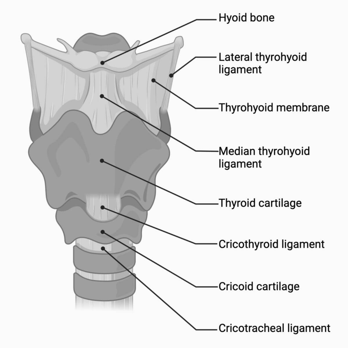
2.5.1. Outer laryngeal ligaments
The four external laryngeal ligaments include:
- the thyrohyoid membrane: quadrangular, between the upper edge of the thyroid cartilage and the hyoid bone
- the median thyrohyoid ligament: median reinforcement of the thyrohyoid membrane
- the lateral thyrohyoid ligament: dorsolateral reinforcement of the thyrohyoid membrane
- the cricotracheal ligament: between the first cartilage clasp of the trachea and the lower edge of the cricoid cartilage
2.5.2. Internal laryngeal ligaments
The internal ligaments of the larynx are collectively referred to as the fibroelastic membrane of the larynx. They include:
- Quadrangular membrane
- Elastic cone
- Vestibular ligament
- Vocal ligament
- Cricothyroid ligament
- Cricoarytenoid ligament
- Cricopharyngeal ligament
- Thyroepiglottic ligament
- Hyoepiglottic ligament
2.6. Vascular supply
The larynx receives its blood supply primarily from two arteries:
- Superior laryngeal artery: a branch of the superior thyroid artery
- Inferior laryngeal artery: a branch of the inferior thyroid artery
2.7. Innervation
The larynx is innervated both motorically and sensory-wise by branches of the vagus nerve, specifically the superior laryngeal nerve and the recurrent laryngeal nerve.
The superior laryngeal nerve provides motor innervation only to the external laryngeal muscle, which is the cricothyroid muscle. The recurrent laryngeal nerve innervates all the internal muscles of the larynx.
For sensory innervation, the glottis serves as the dividing line:
- The superior laryngeal nerve supplies sensation above the glottis.
- The recurrent laryngeal nerve (sometimes referred to as the inferior laryngeal nerve in this context) supplies sensation below the glottis.
3. Embryology
The epithelium of the laryngeal mucosa originates from the endoderm and develops from the laryngotracheal groove. The cartilage of the larynx forms from the mesenchyme of the 4th and 6th pharyngeal (gill) arches. The myoblasts that develop into the laryngeal muscles also arise from these arches.
4. Physiology
The larynx serves several important functions:
- Airway protection
- Voice production (phonation)
- Support during abdominal pressure (e.g., during defecation or lifting)
During swallowing, both the glottis and epiglottis close to prevent food or liquid from entering the lower airways from the mouth or throat.
5. Clinic
5.1. Diseases of the larynx
Diseases affecting the larynx are primarily managed by ENT (Ear, Nose, and Throat) specialists. Common conditions include:
- Laryngitis (inflammation of the larynx)
- Laryngospasm (spasm of the vocal cords)
- Croup (a condition typically in children causing a barking cough)
- Laryngeal fracture
- Laryngeal rupture
- Laryngeal carcinoma (cancer of the larynx)
5.2. Diagnostics
The primary method for examining the larynx is laryngoscopy, which can be complemented with imaging techniques such as MRI when necessary.
5.3. Treatment methods
- Laryngeal surgery
- Laryngeal microsurgery
6. Source
- 3D model: Dr Claudia Krebs (Faculty Lead) University of British Columbia
7. Literature
- Anderhuber et al, Waldeyer - Human Anatomy: Lehrbuch und Atlas in einem Band (19th updated edition), De Gruyter, 2012