Corpus: Trachea


Synonym: windpipe
1. Definition
The term trachea, or windpipe, refers to the tubular structure that connects the larynx to the two main bronchi. It is part of the lower respiratory tract.
2. Anatomy
2.1. Macroscopy
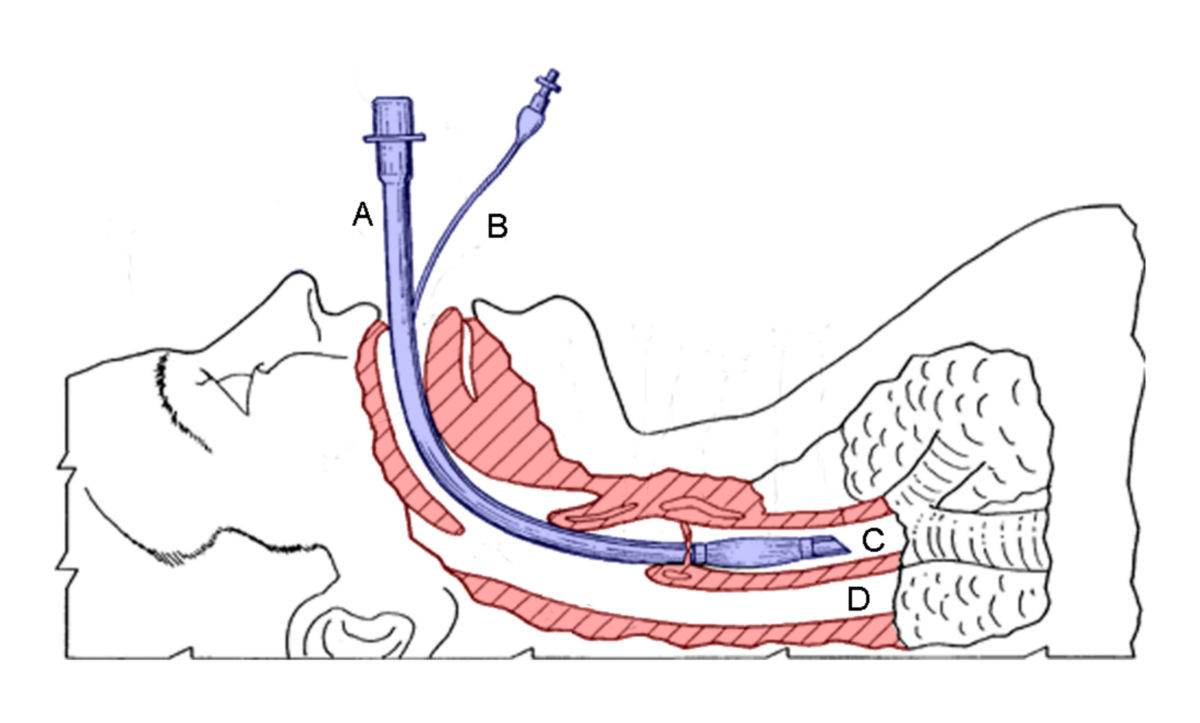
The trachea is approximately 10 to 12 cm long and marks the beginning of the lower respiratory tract. It starts at the cricoid cartilage (ring-shaped cartilage at the base of the larynx) and ends at the tracheal bifurcation, where it divides into the two main bronchi. At this point lies the carina, a ridge that helps guide air into the right and left main bronchi.
The tracheal wall contains 15 to 20 horseshoe-shaped cartilage rings made of hyaline cartilage, connected by ligaments. These cartilage rings support the trachea and prevent it from collapsing during inhalation and exhalation. The average internal diameter of the trachea is 27 mm (sagittal) and 25 mm (coronal) in men, and 23 mm (sagittal) and 21 mm (coronal) in women. The lower limits of normal tracheal diameter are 13 mm in men and 10 mm in women.
2.2. Topography
The trachea is divided into two segments based on its location:
- Cervical Part: This section begins at the lower edge of the cricoid cartilage and extends to the upper opening of the thoracic cavity (thoracic inlet) at the level of the 6th cervical vertebra.
- Thoracic Part: This section runs from the thoracic inlet to the tracheal bifurcation, located at the level of the 4th thoracic vertebra.
In the upper portion of the trachea, the common carotid arteries, internal jugular veins, and vagus nerves lie on either side. These structures are enclosed within the carotid sheath. Anteriorly, the pretracheal layer of cervical fascia covers the trachea, with the thyroid gland situated between this fascia and the trachea in its middle third. Posteriorly, the esophagus lies adjacent to the trachea. The recurrent laryngeal nerves run in the groove between the trachea and the esophagus.
2.3. Histology
The posterior wall of the trachea, known as the membranous wall, lacks cartilage. This region is reinforced by fibroelastic connective tissue and smooth muscle fibers (collectively referred to as the trachealis muscle). The trachealis muscle bridges the open ends of the cartilage rings and can reduce the tracheal lumen by contracting.
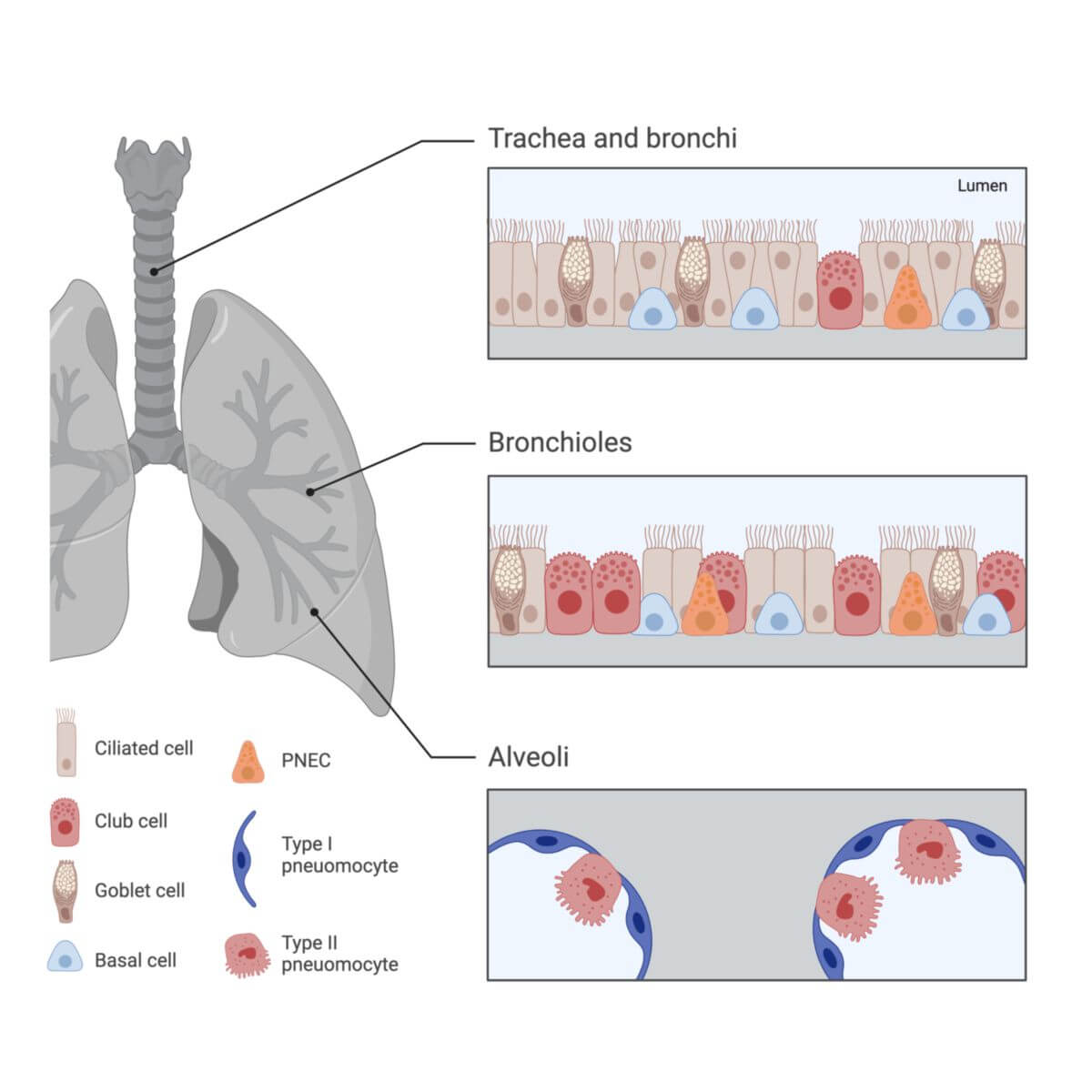
The inner surface of the trachea is lined with respiratory epithelium, which consists of ciliated cells and goblet cells that secrete mucus. Mucus-secreting glands (tracheal glands) are also present in this layer.
The outer surface of the trachea is surrounded by a layer of connective tissue called the tunica adventitia. This layer contains blood vessels and nerves and anchors the trachea to surrounding tissues.
2.4. Blood supply
The blood supply to the trachea is provided by branches from the internal thoracic artery and the inferior thyroid artery, as well as smaller contributions from the superior thyroid artery.
Venous blood from the trachea is drained by the inferior thyroid veins and, to some extent, the esophageal veins, which ultimately empty into the azygos vein (on the right) or the hemiazygos vein (on the left).
2.5. Inntervation
The trachea is innervated by the autonomic nervous system. Sympathetic fibers are derived from the sympathetic trunk, while parasympathetic innervation is provided by branches of the vagus nerve, including the recurrent laryngeal nerve.
3. Clinic
Common medical conditions of the trachea include:
- Tracheitis: Inflammation of the trachea, often associated with infections in other parts of the respiratory tract.
- Williams-Campbell Syndrome: A rare congenital condition characterized by underdevelopment or absence of the tracheal cartilage rings.
- Tracheal stenosis: Narrowing of the trachea, either congenital or acquired, which may require surgical treatment depending on severity.
- Tracheal diverticula: Outpouchings of the tracheal wall. These are uncommon and usually asymptomatic.
If the tracheal index (ratio of the coronal diameter to the sagittal diameter) is less than 0.6, the condition is referred to as "saber trachea," while a ratio greater than 1.0 is called "discoid trachea."