Corpus: Mylohyoid muscle


from ancient Greek: μύλη ("myle") - millstone, mill tooth; ὑοειδής ("hyoides") - υ-shaped
1. Definition
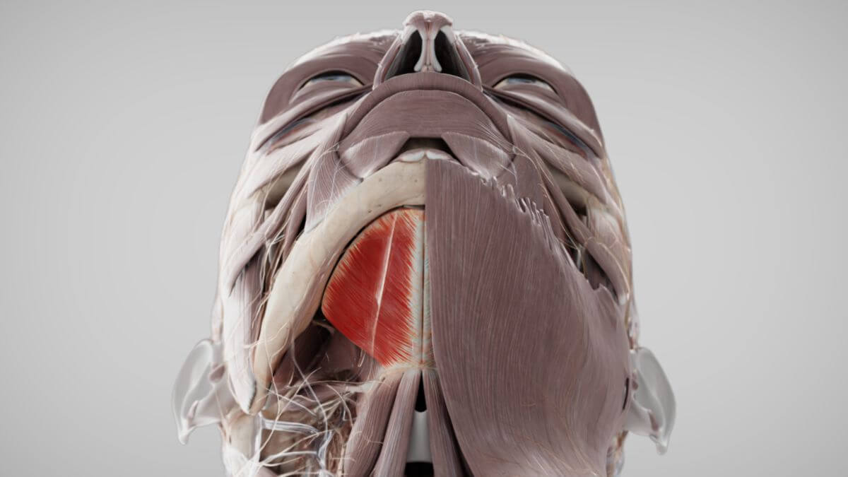
The mylohyoid muscle is a skeletal muscle that is part of the suprahyoid musculature, forming the muscular foundation of the floor of the mouth. It is derived from the first branchial arch.
2. Course
The mylohyoid muscle originates from the mylohyoid line (linea mylohyoidea) on the inner surface of the mandible. From its origin, the muscle fibers run downward towards the hyoid bone. The posterior fibers of the mylohyoid muscle attach directly to the hyoid bone, while the anterior fibers converge with those from the opposite side at the midline connective tissue structure called the mylohyoid raphe, which extends from the chin to the hyoid bone.
3. Innervation
The mylohyoid muscle is innervated by the mylohyoid nerve, a branch of the inferior alveolar nerve, which is itself a branch of the mandibular nerve (cranial nerve V3).
4. Vascular supply
The muscle receives its blood supply from the mylohyoid branch of the inferior alveolar artery.
5. Function
The mylohyoid muscle plays a key role in the swallowing process by pulling the hyoid bone anteriorly. It also contributes to jaw opening and assists in the grinding movement of the jaw. As a broad muscle, the mylohyoid forms the muscular floor of the mouth, also known as the diaphragm oris.
6. Etymology
The term "mylohyoid muscle" reflects its anatomical course, indicating both its origin and insertion points. The muscle originates from the mandible in the area of the molars (derived from the ancient Greek word "mylai" meaning molars) and extends to the hyoid bone (os hyoideum).