Corpus: Lacrimal bone


1. Definition
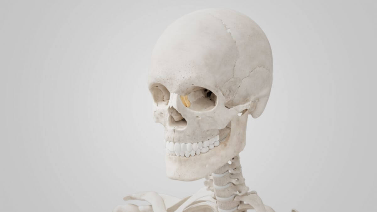
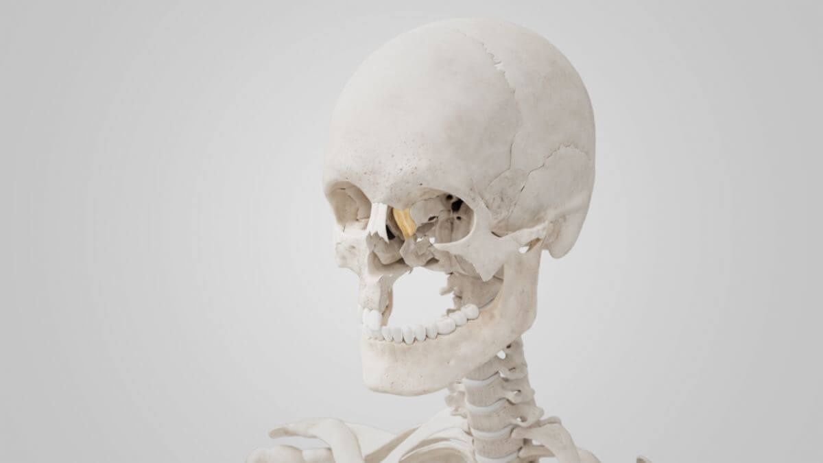
2. Anatomy
The lacrimal bone has two surfaces and four margins.
2.1. Lateral surface
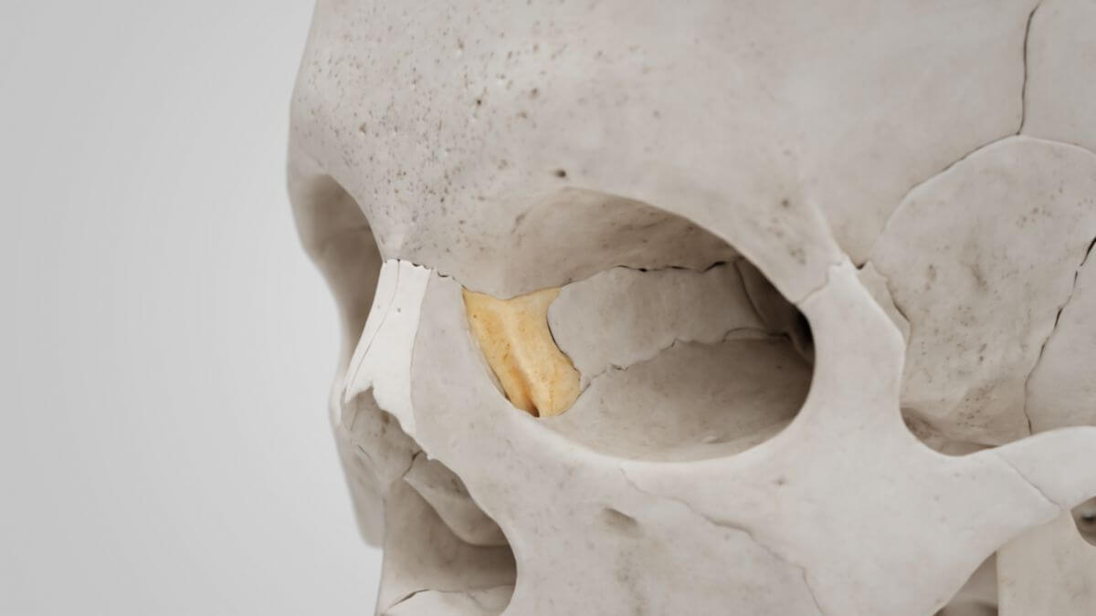
The lateral or orbital surface of the lacrimal bone is divided into two halves by a bony crest, the posterior lacrimal crest. In front of the crest is the lacrimal groove, the inner edge of which joins the frontal process of the maxilla, completing the lacrimal fossa. The upper part of this fossa harbors the lacrimal sac, while the lower part contains the beginning of the nasolacrimal duct.
The surface behind the crista lacrimalis is smooth and contributes to the formation of the medial orbital wall. The crista itself serves as the origin for the lacrimal part of the orbicularis oculi muscle. It ends in a small, hook-like projection, the lacrimal hamulus, which articulates with the lacrimal tubercle of the maxilla, forming the superior opening of the nasolacrimal canal.
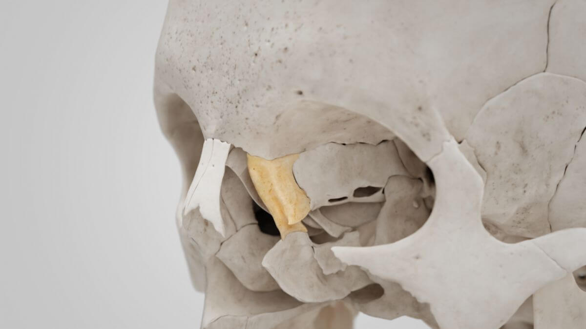
2.2. Medial surface
The medial or nasal surface of the lacrimal bone has a bony groove that corresponds with the posterior lacrimal crest. The bony surface anterior to the groove comprises a part of the middle nasal meatus. Posterior to the groove, the bone articulates with the ethmoid bone and completes some anterior ethmoid cells.
2.3. Margins
The margins of the lacrimal bone are in contact with four other cranial bones. The anterior margin articulates with the frontal process of the maxilla. The posterior margin articulates with the orbital plate of the ethmoid bone. The superior margin unites with the frontal bone. The inferior margin articulates partly with the maxilla and partly with the inferior nasal concha.