Corpus: Lacrimal gland


Synonym: tear gland, glandula lacrimalis
1. Definition
The lacrimal gland is part of the lacrimal system and is responsible for producing tears. It is located in the upper outer corner (temporal side) of the eye socket, embedded in a depression called the lacrimal fossa of the frontal bone.
2. Anatomy
The lacrimal gland is divided by the tendon of the superior levator palpebrae muscle into two parts: the orbital part, located above the tendon and the papebral part, located below the tendon. The lacrimal gland consists of several lobes separated from each other by connective tissue. The orbital part is also referred to as Galen's gland, named after Galen of Pergamon.
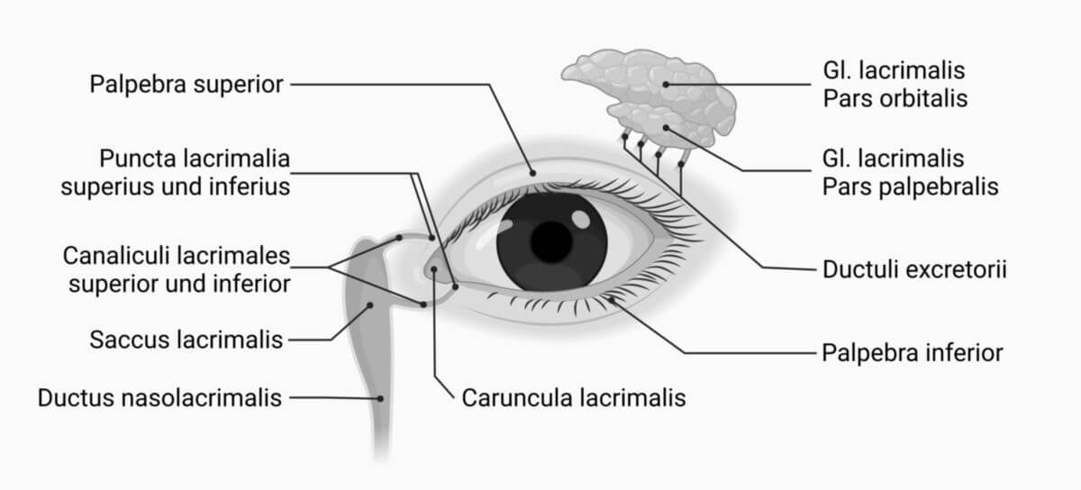
In addition to the main lacrimal gland, there are smaller accessory lacrimal glands found in the upper eyelid:
2.1. Innervation
The lacrimal gland receives autonomic innervation via the lacrimal nerve, which includes parasympathetic and sympathetic nerve fibers from different sources.
2.1.1. Parasympathetic innervation
Parasympathetic fibers originate from the facial nerve (cranial nerve VII), specifically from its intermediate nerve. These fibers exit the facial nerve at the hiatus of the greater petrosal nerve and travel through the pterygoid canal to reach the pterygopalatine ganglion, where they synapse. Postganglionic fibers then join the zygomatic nerve and eventually merge with the lacrimal nerve in the lateral wall of the orbit, delivering parasympathetic signals to the lacrimal gland.
Some sources also suggest that orbital branches of the pterygopalatine ganglion contribute to the innervation of the lacrimal gland.
2.1.2. Sympathetic innervation
Sympathetic fibers originate from the internal carotid plexus and travel via the deep petrosal nerve. These fibers join with the greater petrosal nerve in the pterygoid canal to form the nerve of the pterygoid canal. Unlike parasympathetic fibers, the sympathetic fibers pass through the pterygopalatine ganglion without synapsing and then follow the same path to the lacrimal gland.
2.2. Blood supply
The lacrimal gland receives its blood supply from the lacrimal artery, a branch of the ophthalmic artery. Venous blood is drained via the lacrimal vein into the superior ophthalmic vein.
3. Histology
The lacrimal gland is a tubulo-alveolar gland that produces only serous (watery) secretions. Some sources also describe it as a tubulo-acinous gland, while others include both terms. The glandular tissue is divided into distinct lobes by connective tissue. Tears are secreted via up to 10 excretory ducts, which empty into the conjunctival fornix and then flow into the lacrimal drainage system.
To distinguish the lacrimal gland from other purely serous glands, such as the parotid gland or the exocrine pancreas, note that the lacrimal gland lacks the striated ducts and intercalated ducts present in those glands.
4. Clinic
Inflammation of the lacrimal gland is called dacryoadenitis.