Corpus: Trigeminal nerve


Synonyms: 5th cranial nerve, V nerve, triplet nerve
1. Definition
The trigeminal nerve is the fifth cranial nerve and carries general sensory and specific motor fibers. It transmits sensory information from the entire facial area to the brain and innervates the masticatory muscles.
2. Embryology
The trigeminal nerve is classified as a branchial arch nerve and develops from the nerve of the first branchial arch. It innervates all muscles derived from the muscle anlage of this arch.
3. Course
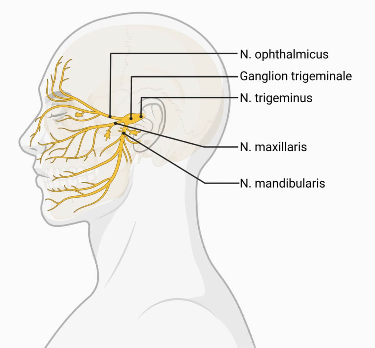
The trigeminal nerve originates from the lateral edge of the pons and runs to the petrous part of the temporal bone, where it pierces the dura mater. At its origin, it has two roots:
- Sensory root (radix sensoria)
- Motor root (radix motoria)
The trigeminal nerve swells into the trigeminal ganglion (Gasserian ganglion) in a dural pocket above the foramen lacerum, the trigeminal cave. It then divides into its three terminal branches: the ophthalmic nerve, the maxillary nerve, and the mandibular nerve.
3.1. Ophthalmic nerve (V1)
The ophthalmic nerve enters the cavernous sinus and runs forward in its lateral wall. Before it enters the orbit through the superior orbital fissure, it gives off a recurrent meningeal branch to the meninges. On entering the orbit, it splits into its three terminal branches: the frontal nerve, nasociliary nerve, and lacrimal nerve.
3.2. Maxillary nerve (V2)
The maxillary nerve runs forward in the basolateral wall of the cavernous sinus, where it gives off a meningeal branch before passing through the foramen rotundum. It reaches the pterygopalatine fossa via the foramen rotundum and divides into its terminal branches: the zygomatic nerve, infraorbital nerve, and ganglionic branches.
3.3. Mandibular nerve (V3)
The mandibular nerve passes through the foramen ovale into the infratemporal fossa. Unlike the other two branches of the trigeminal nerve, the mandibular nerve gives off a recurrent meningeal branch outside the cranial cavity. Together with the middle meningeal artery, it re-enters the cranial cavity via the foramen spinosum to supply sensation to the meninges. In the infratemporal fossa, it divides into four general sensory terminal branches:
and the special motor branches that innervate the masticatory muscles. Some of these fibers are also referred to as the masticatory nerve. The mandibular nerve is the only branch of the trigeminal nerve that contains motor fibers.
4. Fibre qualities
The sensory fibers of the sensory root (radix sensoria) are known as the portio major nervi trigemini after the trigeminal ganglion. The motor fibers from the motor root (radix motoria) pass through the trigeminal ganglion and join the mandibular nerve, forming the smaller part known as the minor portion nervi trigemini.
The major portion is purely sensory. It contains fibers of epicritical, protopathic, and proprioceptive sensitivity, with their nerve cell bodies predominantly located in the trigeminal ganglion. The nerve cell bodies of the proprioceptive neurons are located in the mesencephalic nucleus of the trigeminal nerve in the brainstem for developmental reasons.
The mixed minor portion consists mainly of motor fibers. It contains a small proportion of proprioceptive fibers that run to the muscles it supplies. However, these fibers also originate from the sensory root.
5. Innervation
5.1. Ophthalmic nerve
The ophthalmic nerve supplies the entire orbit, the skin of the forehead and nose, part of the paranasal sinuses, and the nasal septum mucosa. One of its branches, the lacrimal nerve, carries parasympathetic fibers from the facial nerve to the lacrimal gland.
5.2. Maxillary nerve
The maxillary nerve innervates almost the entire nasal cavity mucosa, the palate, the upper jaw with gums and teeth, the skin between the lower eyelid and upper lip, and part of the temporal region.
5.3. Mandibular nerve
The mandibular nerve supplies sensation to the skin over the chin up to the temple, the front two-thirds of the tongue, and the lower jaw with teeth and gums. Through its special motor fibers, it innervates the masticatory muscles, the tensor tympani muscle, the tensor veli palatini muscle, the mylohyoid muscle, and the anterior belly of the digastric muscle. The motor fibers of the trigeminal nerve run exclusively in the mandibular nerve.
Additionally, branches of the auriculotemporal nerve and lingual nerve carry parasympathetic fibers from the glossopharyngeal nerve to the parotid gland and from the facial nerve to the submandibular and sublingual glands.
6. Core areas
6.1. General somatosensory fibres
6.1.1. Mesencephalic nucleus of the trigeminal nerve
Located in the midbrain, it receives afferent fibres from muscle spindles of the masticatory muscles and sends efferent fibres to them. This nucleus is composed of pseudounipolar nerve cells, and the afferents do not lead to the trigeminal ganglion but directly to the cranial nerve nucleus.
6.1.2. Principal sensory nucleus of the trigeminal nerve
Located in the pons, it receives fibers responsible for epicritical sensibility from the face.
6.1.3. Spinal nucleus of the trigeminal nerve
Located in the medulla oblongata and spinal cord, it receives fibers of protopathic sensitivity from the face. It can be subdivided into pars caudalis, pars interpolaris, and subnucleus oralis, explaining the bowl-shaped, perioral innervation areas (Sölder lines).
6.2. Special visceromotor fibres
6.2.1. Spinal accessory nucleus
Located in the pons, it provides motor innervation to the muscles derived from the first branchial arch.
7. Clinic
7.1. Diseases
A lesion of the trigeminal nerve results in loss of sensitivity and/or paralysis of the supplied regions. Depending on the lesion's location, sensory loss can be central or peripheral. Other diseases of the trigeminal nerve include:
- Trigeminal neuralgia
- Trigeminal neurinoma
- Herpes zoster ophthalmicus
- Nevus of Ota
7.2. Diagnostics
Irritations in the innervation areas of the three main branches can be diagnosed via the trigeminal pressure points (Valleix points):
- Supraorbital foramen for the supraorbital nerve (ophthalmic nerve)
- Infraorbital foramen for the infraorbital nerve (maxillary nerve)
- Mental foramen for the mental nerve (mandibular nerve)
These pressure points are painful when the corresponding branch is diseased, particularly in trigeminal neuralgia.