Corpus: Cranial nerve


1. Definition
Cranial nerves are nerves whose fibers either emerge directly from the brain or extend into it. This differentiates them from spinal nerves, which arise from the spinal cord. Most cranial nerves are connected to specialized collections of nerve cells in the brainstem, known as cranial nerve nuclei. Each cranial nerve passes through at least one opening in the skull's bony structure.
2. Fiber qualities
Half of the cranial nerves are mixed nerves, meaning they carry fibers with multiple functions, such as controlling muscles while simultaneously transmitting sensory information. The other half carries only one type of fiber. The fiber types are divided into:
2.1. Efferent fibers
- General somatic efferent (GSE): control voluntary muscle movement.
- General visceral efferent (GVE): parasympathetic, regulate smooth muscle, cardiac muscle, and glands.
- Special visceral efferent (SVE): control muscles derived from embryonic gill arches
2.2. Afferent fibers
- General somatic afferent (GSA): transmit sensations such as touch, pain, and temperature from skin and mucosa.
- Special somatic afferent (SSA): involved in vision, hearing, and balance
- General visceral afferent (GVA): carry information from internal organs
- Special visceral afferent (SVA): responsible for taste and smell
"General" fibers are found throughout the peripheral nervous system, while "special" fibers are unique to cranial nerves and are associated with specialized functions such as sensory perception or innervation of gill-arch-derived muscles.
3. Systematics
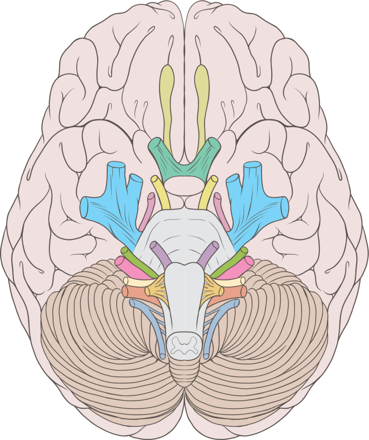
The 12 cranial nerves are numbered with Roman numerals in the order in which they emerge from the brain, from front (rostral) to back (caudal). This classification was introduced in 1788 by Samuel Thomas von Soemmerring.
The olfactory nerve (I) consists of fibers (fila olfactoria) that connect to the olfactory bulb. Occasionally, the rudimentary Jacobson’s organ in humans is referred to as the 1st cranial nerve. The optic nerve (II) transmits signals from the retina to the brain.
Modern consensus considers the 1st and 2nd cranial nerves as extensions of the brain rather than true nerves, although they are still classified as cranial nerves.
The accessory nerve (XI) is also unique because some of its fibers originate in the spinal cord.
| Nervus | Name' | Function | fiber qualities |
|---|---|---|---|
| I | Olfactory nerve | Conducts signals from the nose to the brain | SVA |
| II | Optic nerve | Conducts the signals from the retina to the brain | SSA |
| III | Oculomotor nerve | Controls eye movements, the eyelid retractor and the iris | GSE, GVE |
| IV | Trochlear nerve | Controls the superior oblique muscle of the eye | GSE |
| V * | Trigeminal nerve | Subdivided into the ophthalmic nerve, the maxillary nerve and the mandibular nerve. It transmits sensitive information from the entire facial area to the brain and innervates the masticatory muscles. | GSA, SVE |
| VI | Abducens nerve | Innervates the lateral rectus muscle of the eye | GSE |
| VII * | Facial nerve | Controls the muscles of facial expression and the stapedius muscle. It also mediates the perception of taste in the front two thirds of the tongue, innervates all head glands except the parotid gland | SVE, GVE, GSA, SVA |
| VIII | vestibulocochlear nerve | Responsible for transmitting information from the cochlea and the organ of balance | SSA |
| IX * | Glossopharyngeal nerve | Conducts the signals from the posterior part of the tongue to the brain and innervates the muscles of the pharynx. Important for the act of swallowing. Also innervates the parotid gland. | GSA, GVE, SVE, GVA, SVA |
| X * | Vagus nerve | Main nerve of the parasympathetic nervous system and involved in regulating the activity of many internal organs | GSA, GVE, SVE, GVA, SVA |
| XI (*) | Accessory nerve | Supplies the trapezius and sternocleidomastoid muscles | GSE, (SVE) |
| XII | Hypoglossal nerve | Controls the movement of the tongue | GSE |
| *) embryological: gill arch nerves | |||
The cranial nerves V, VII, IX and X are also classified as gill arch nerves due to their embryonic developmental history. Their motor fiber qualities are described as special visceromotor or branchiomotor. They supply muscles that have developed from the muscle anlage of the gill arches.
In some textbooks, nerve XI (nervus accessorius) is also categorised as a gill arch nerve. However, this only applies to its cranial part ("internal ramus"), which is a continuation of the vagus nerve (nerve X).
The 7th cranial nerve (facial nerve) has an occasionally separate component, the nervus intermedius, which is sometimes called the "13th cranial nerve." This is relevant when studying the parasympathetic system of the head.
Regarding the 7th cranial nerve: its classification is also not standardised. Sometimes a part of the 7th cranial nerve, the intermediate nerve, is referred to as the "13th cranial nerve". This concept is useful for understanding the function of the parasympathetic nervous system in the head area.
In addition to the 12 cranial nerves mentioned above, the terminal nerve, which was only discovered in 1913, can also be counted among the cranial nerves (nerve 0).
4. Course
All cranial nerves are arranged in pairs. After emerging from the nerve cell mass of the brain, the fibers of the cranial nerves initially run intracranially and then emerge from the skull via differently dimensioned channels (foramina, fissures). Their extracranial section then begins.
| Nerve | Passage |
|---|---|
| Olfactory nerve (I) | Cribriform plate |
| Optic nerve (II) | Optic canal |
| Oculomotor nerve (III), trochlear nerve (IV), ophthalmic nerve (V1), abducens nerve (VI) | Superior orbital fissure |
| Maxillary nerve (V2) | Foramen rotundum |
| Mandibular nerve (V3) | Foramen ovale |
| Facial nerve (VII) | Facial canal |
| Vestibulocochlear nerve (VIII) | Internal acoustic meatus |
| Glossopharyngeal nerve (IX), vagus nerve (X), accessory nerve (XI) | Jugular foramen |
| Hypoglossal nerve (XII) | Hypoglossal canal |
5. Ganglia
Cranial nerves with sensory components (V, VII, VIII, IX, X) contain cell bodies in ganglia located outside the brain. These cranial nerve ganglia correspond to the dorsal root ganglia of the spinal nerves. These include:
- Trigeminal ganglion (trigeminal nerve)
- Geniculate ganglion (facial nerve)
- Cochlear ganglion (vestibulocochlear nerve)
- Vestibular ganglion (vestibulocochlear nerve)
- Superior and inferior ganglion of the vagus nerve
- Superior and inferior ganglion of the glossopharyngeal nerve
Some cranial nerves also carry fibers from nerve cells in the parasympathetic head ganglia:
- Ciliary ganglion (oculomotor nerve)
- Pterygopalatine ganglion (maxillary nerve)
- Ganglion oticum (glossopharyngeal nerve)
- Submandibular ganglion (lingual nerve)
6. Preparations
Brain stem with cranial nerves
Cranial base with cranial nerves
7. Clinic
Damage to cranial nerves is called cranial nerve palsy. Causes include trauma, infections, tumors, or ischemia. The damage can occur centrally (in the nucleus) or peripherally (along the nerve’s course). Examples include facial nerve palsy and oculomotor nerve palsy.
Pain syndromes, such as trigeminal neuralgia, glossopharyngeal neuralgia, or intermedius neuralgia, are associated with sensory cranial nerve dysfunction.
8. Mnemonics
Mnemonic for the 12 fiber qualities of the cranial nerves (s=sensitive; m=motor; b=both):
- "Some say money matters, but my brother says big boobs matter more."
- "Some students make money, but my brother says Boris Becker makes more,"
9. Sources
- 3D model: Dr. Claudia Krebs (Faculty Lead) University of British Columbia