Corpus: Occipitofrontalis muscle


1. Definition
The occipitofrontalis muscle, part of the epicranial muscles, consists of two muscle bellies located at opposite ends of the skull, connected by the galea aponeurotica. It is classified as part of the mimic musculature.
2. Course
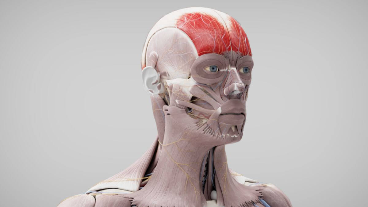
The frontal belly of the muscle originates at the supraorbital margin of the frontal bone and the area around the glabella. Its fibers extend into neighboring facial muscles, including the procerus, corrugator supercilii, and orbicularis oculi muscles.
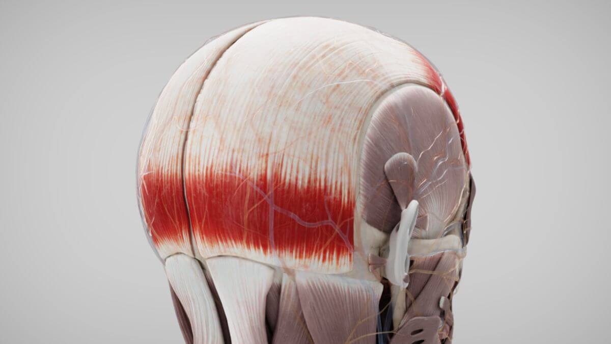
The occipital belly originates from the highest nuchal line of the occipital bone and, to a lesser extent, from the temporal bone.
The fibers of both muscle bellies run vertically upward and insert into the galea aponeurotica, the tendinous sheet covering the top of the skull.
3. Morphology
Both muscle bellies have an almost quadrangular shape. The frontal belly of the occipitofrontalis muscle is more prominent and contains longer fibers.
4. Innervation
The frontal belly of the occipitofrontalis muscle is innervated by the temporal branches of the facial nerve, while the occipital belly is innervated by the posterior auricular nerve, also a branch of the facial nerve.
5. Function
6. Clinic
Due to the tension exerted by the occipitofrontalis and temporoparietalis muscles on the galea aponeurotica, scalp injuries often result in gaping wounds.