Corpus: Tarsus (foot)


Synonym: tarsal bones
1. Definition
The tarsus is located between the malleolar fork of the lower leg and the metatarsus. The bones in this region belong to the short bones.
In the eye, "tarsus" is used to describe the palpebral tarsus, a dense connective tissue structure within the eyelid.
2. Anatomy
The tarsus consists of two rows of tarsal bones. These bones are connected through joints and are tightly stabilized by ligaments, which maintain their structural relationships.
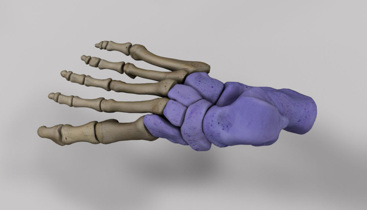
The 7 tarsal bones are divided into a proximal and a distal row. The proximal row articulates with the fibula and tibia, the distal row is adjacent to the metatarsal bones of the forefoot. The joint line between the proximal and distal row is called the Chopart joint line.
2.1. Proximal row
2.2. Distal row
- Navicular bone
- Medial, intermediate, and lateral cuneiform bones (ossa cuneiformia I - III)
- Cuboid bone
2.3. Joints
The tarsal bones form the following joints:
- Talocrural joint: Talus, fibula and tibia
- Talotarsal joint:
- Subtalar joint: Calcaneus and talus
- Talocalcaneonavicular joint: Talus, calcaneus, navicular bone
- Transverse tarsal joint (Chopart joint):
- Calcaneocuboid joint: Calcaneus and cuboid bone
- Talonavicular joint: Talus and navicular bone
- Cuneonavicular joint: Navicular bone and cuneiform bones
- Joints between the distal tarsal bones:
- Intercuneiform joints between cuneiform bones
- Cuneocuboid joint: Lateral cuneiform and cuboid bones
- Tarsometatarsal joint (Lisfranc joint): Distal tarsal bones and metatarsals
Unlike the upper and lower ankle joints, the other joints in the tarsus have limited mobility (amphiarthroses), as they are stabilized by strong ligament connections. These joints move primarily passively when the foot makes contact with the ground, allowing the foot to act as a flexible, spring-like platform that adapts to uneven terrain. Combined, the limited mobility of individual joints allows for overall torsion of the foot, which is essential during gait and weight-bearing activities.
3. Function
The tarsus transmits forces between the forefoot and the lower leg. The subtalar joint enables complex three-dimensional movements, allowing the foot to perform eversion (pronation) and inversion (supination).
In contrast, the transverse tarsal joint (Chopart joint) permits movement along two axes. Both joint systems work together to either increase the flexibility of the foot or stiffen it, depending on the position and activity. For example, eversion increases midfoot flexibility, while inversion stabilizes it.
4. Clinic
In clinical practice, the tarsus is divided functionally rather than anatomically. The nomenclature differs from the anatomical designations.
- The two proximal tarsal bones (talus and calcaneus) are referred to as the hindfoot.
- The five distal tarsal bones are grouped with the metatarsals as the midfoot.
- The toes and their associated metatarsals are categorized as the forefoot.
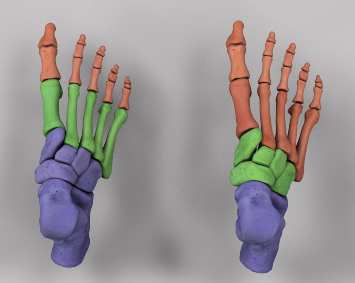
- Left: Anatomical classification of the foot skeleton. Blue - tarsus, green - metatarsus, red - antetarsus
- Right: Clinical anatomy of the foot skeleton. Blue - hindfoot, green - metatarsus, red - forefoot