Corpus: Cuboid bone


2. Anatomy
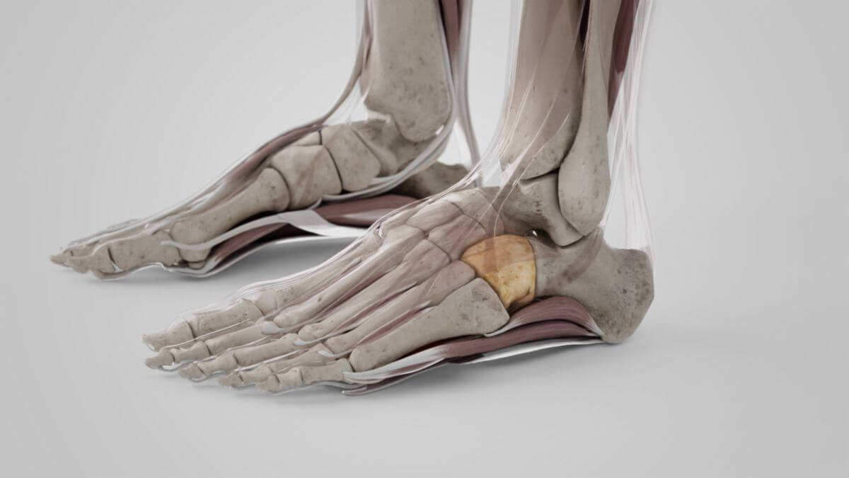
2.1. Location
The cuboid bone lies on the lateral side of the foot, in front of the calcaneus and behind the metatarsal bones IV and V. It articulates with four neighboring bones: the calcaneus, the cuneiform bone III and the metatarsal bones IV and V. Occasionally it also articulates with the navicular bone.
2.2. Surfaces
2.2.1. Dorsal surface
The dorsal surface of the cuboid bone is inclined cranially and laterally. It is rough and serves as an attachment for some ligaments.
2.2.2. Plantar surface
The surface of the bone facing the plantar side of the foot shows a distinct depression at the front, the peroneal sulcus, which runs obliquely to the front and medially. It accommodates the tendon of the fibularis longus muscle and is bordered at the rear by a prominent bony ridge, the tuberositas ossis cuboidei, to which the long plantar ligament attaches. This bony ridge ends laterally in a small elevation, the surface of which has an oval facet. It is in direct contact with the sesamoid bone, which is embedded in the tendon of the fibularis longus muscle. The bone surface behind the sulcus is rough. This is where the calcaneocuboid plantar ligament and some tendon fibres of the posterior tibialis muscle attach. The surface also serves as the origin for some fibres of the flexor hallucis brevis muscle.
2.2.3. Posterior surface
The posterior surface of the bone is smooth, triangular and concave-convex. It articulates with the calcaneus.
2.2.4. Anterior surface
The anterior surface of the cuboid bone is smaller than the posterior surface, but is also triangular in shape. It is divided into two articular facets by a ridge. The smaller medial facet is quadrangular and articulates with metatarsal bone IV, the larger lateral surface with metatarsal bone V.
2.2.5. Lateral surface
The lateral side of the cuboid bone facing the outer edge of the foot is slightly concave and serves as a pulley for the tendon of the peroneus longus muscle.
2.2.6. Medial surface
The joint surface for the neighboring cuneiform bone III is located on the medial surface, facing the inner edge of the foot.
3. Development
The bone core of the cuboid bone usually appears in the 10th fetal month (sign of maturity).