Corpus: Cuneiform bone

1. Definition
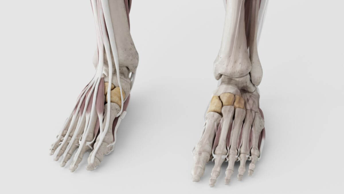
The three cuneiform bones are bones that belong to the distal row of tarsal bones. They are located between the navicular bone and the metatarsal bones I-III.
2. Anatomy
The three cuneiform bones are small and compact and belong to the short bones. They are numbered systematically from medial to lateral:
- cuneiform bone I (medial)
- cuneiform bone II (intermedial)
- cuneiform bone III (lateral)
3. Joints
The cuneiform bones articulate proximally, i.e. towards the tarsus, with the navicular bone and form with it the cuneonavicular joint. Distally, they articulate with the metatarsal bones I-III and are thus involved in the formation of the tarsometatarsal joint. These joints form the medial part of the Lisfranc joint line. The cuneiform bones are connected to each other by two intercuneiform joints. The lateral cuneiform bone and the cuboid bone also form the cuneocuboid joint.
4. Ligaments
The above-mentioned joint connections are secured by numerous ligaments. These include, among others:
5. Development
The cuneiform bones develop from the 5th week of development from mesenchymal condensations in which the cells differentiate into chondroblasts. Ossification only takes place postnatally — at birth the bones still consist entirely of cartilage. The bone nuclei of the cuneiform bones appear at different times:
- medial cuneiform bone: in the 2nd to 3rd year of life
- intermedial cuneiform bone: in the 3rd year of life
- lateral cuneiform bone: in the 1st to 2nd year of life
6. Clinic
The cuneiform bones can be damaged in the context of tarsal fractures in isolation or in conjunction with other bones. As they are important for the geometry of the foot, surgical fracture treatment is often necessary.