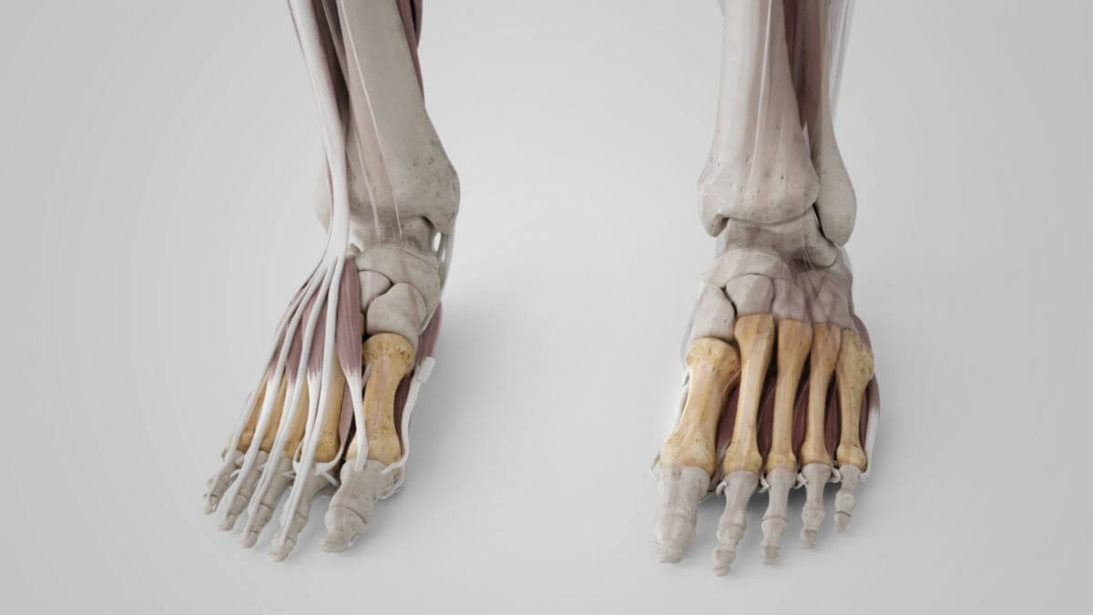
Corpus: Metatarsal bone


1. Definition
The 5 metatarsal bones form the bony base of the metatarsus.
2. Classification
The metatarsal bones are simply numbered systematically from medial to lateral for their correct anatomical designation, with metatarsal bone I carrying the big toe:
- metatarsal bone I
- metatarsal bone II
- metatarsal bone III
- metatarsal bone IV
- metatarsal bone V
The intermetatarsal spaces are located between the bones.
3. Anatomy
3.1. Common features
All metatarsal bones are long bones consisting of three sections. From proximal to distal, these are:
- Base (also: extremitas tarsalis or proximalis)
- Shank (corpus)
- Head (capitulum or caput, also: extremitas phalangealis or distalis)
The bases of the metatarsals have a wedge-shaped basic form. Their concave cartilage surfaces articulate with the tarsal bones in the form of the tarsometatarsal joints. They are also connected to each other by 4 intermetatarsal joints, some of which communicate with the neighboring tarsometatarsal joints.
The shaft has a triangular cross-section, with the tip pointing towards the plantar side. The plantar bone edge is concavely curved in the longitudinal direction, the dorsal surface is straight to slightly convex.
The head has a convex wedge-shaped cartilaginous surface that extends further proximally on the plantar side. There is a small tubercle on both sides of the head, to which the collateral ligaments of the metatarsophalangeal joint attach.
3.2. Differences
Metatarsal bone I is the shortest and strongest metatarsal bone. Its larger cross-section is due to the increased biomechanical load caused by the rolling movement of the big toe. Metatarsal bone II is the longest metatarsal bone. The length of the following metatarsal bones decreases continuously laterally.
4. Development
The initially cartilaginous metatarsal bones receive perichondral bone cuffs in the area of the corpus during the 2nd and 3rd fetal month. There is also an epiphyseal bone attachment in each case. The similarity to the metacarpal bones can be recognised from the ossification: the first metatarsal bone has its epiphyseal bone core at the base, the other metatarsal bones in the head. The epiphyseal enchondral bone anlagen do not appear until the 2nd to 4th year of life. In rare cases, an additional, second epiphyseal anlage can be observed in the first and fifth metatarsal bone.
5. Biomechanics
Despite the numerous ligamentous structures that connect them to each other, the metatarsal bones can be moved slightly against each other. The slight spreading and fanning out allows the forefoot to be deformed, which comes into play during pronation and supination and can better compensate for unevenness of the ground.
6. Clinic
A pathological splaying of the metatarsal bone I medially is referred to as metatarsus primus varus. It often occurs together with hallux valgus. To determine the severity of the deformity, the angle between metatarsals I and II (intermetatarsal angle) is measured on the X-ray. It is normally a maximum of 8 to 10°.