Corpus: Tibia

Synonym: shinbone
1. Definition
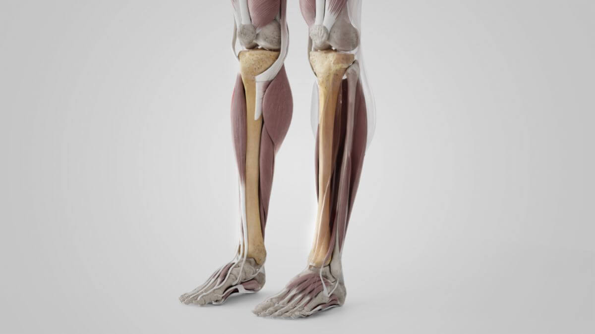
The tibia is the second-longest bone of the human skeleton after the femur. It can be roughly divided into the tibial shaft (corpus tibiae) and the two bone sections involved in the knee and ankle joint (extremitas proximalis and extremitas malleolaris).
2. Anatomy
2.1. Extremitas proximalis
The extremitas proximalis — also known as the head of tibia — is a large and massive section of bone that is made up of two large bony bulges, the condylus medialis and the condylus lateralis. Cranially, the structures are covered with two concave cartilage facets.
The medial facet has an oval basic shape, the lateral facet is almost round. The central parts of both articular surfaces articulate with the two femoral condyles, the peripheral parts are in contact with the menisci. The eminentia intercondylaris is located between the articular surfaces, with a prominent tubercle on each side, onto which the cartilage surfaces extend. Dorsal and ventral to the intercondylar eminence are roughened bone depressions, the anterior tibial intercondylar fossa and the posterior tibial intercondylar fossa. The fibers of the anterior and posterior cruciate ligaments and the medial meniscus radiate into these depressions.
2.2. Corpus tibiae
The tibial shaft (corpus tibiae) has a triangular cross-section and accordingly has three surfaces (facies) and three edges (margins).
2.2.1. Anterior border
The anterior border, also known as the crista anterior, is the edge of the bone pointing forwards. It begins at the tibial tuberosity and ends distally at the anterior edge of the medial malleolus. In the upper part it protrudes as a clear edge, in the lower part it is smoother and rounded. It serves as an attachment for the deep leaf of the tibial fascia.
2.2.2. Medial border
The medial border is more rounded at the top and bottom, but is more prominent in the center. It begins proximally at the back of the medial condyle and ends distally at the posterior edge of the medial malleolus. Its upper parts serve as an attachment for the tibial collateral ligament. Some fibers of the popliteus muscle also insert here. Fibers of the soleus and flexor digitorum longus muscles originate in the middle third.
2.2.3. Lateral border
The lateral border or crista interossea is a thin, protruding bony edge that serves as an attachment for the interosseous membrane. It begins proximally in front of the lateral articular surface and divides into two lamellae further distally.
2.3. Extremitas distalis
The extremitas malleolaris (also: extremitas distalis) is significantly smaller than the extremitas proximalis. A distinction can be made between 5 surfaces. On the medial side it has a pronounced protrusion, the malleolus medialis. Due to its shape, this section of the tibia is also known as the pilon tibiale.
3. Development
Perichondral ossification of the corpus tibiae begins during the 7th week of fetal life. In the 10th fetal month or 1st year of life, an enchondral bone nucleus appears at the proximal end, whereas the distal epiphysis does not receive its enchondral bone nucleus until the beginning of the 2nd year of life. Closure of the epiphyses occurs distally during the 17th and 19th year of life, proximally only between the 19th and 20th year of life.
4. Clinic
On X-ray images, epiphyseal joints, especially in the area of the distal epiphysis, can be confused with fracture lines.