Corpus: Femur


Synonym: thighbone
1. Definition
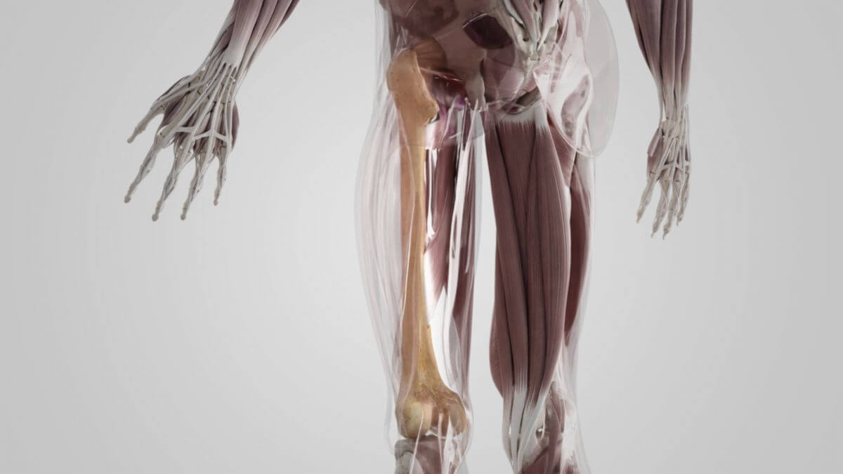
2. Structure
The largest part of the femur is formed by the body of the femur (corpus femoris). It has a triangular base in cross-section and accordingly has three edges. At the proximal end of the femur is the head of the femur (caput femoris), which rests on a femoral neck (collum femoris) that is inclined at an angle of around 135° to the axis of the femur. The angle between the neck and shaft is known as the collum-diaphyseal angle (CD angle). At the lateral end of the connection between the neck and the femoral shaft, there are two bony protrusions known as trochanters:
- the greater trochanter on the ventral side, at the craniolateral edge of the femoral shaft and
- the lesser trochanter on the dorsal side. Compared to the greater trochanter, it is located further distally and medially.
Both trochanters serve, among other things, as attachment points for the thigh muscles. The same applies to the intertrochanteric line located between the two trochanters, which connects them on the ventral side of the femur. On the dorsal side there is a sharp bone ridge, the intertrochanteric crest. The linea aspera runs along the dorsal femoral shaft and serves as the attachment for almost all femoral adductors. Distally, the linea aspera is divided into two bone ridges, the medial supracondylar line and the lateral supracondylar line. Together with the intercondylar line, they frame a triangular surface, the popliteal surface. The distal end of the femur is occupied by the two articular cartilages (femoral condyles) opposite the tibia, which are known as the lateral and medial condyle. The depression between the condyles is referred to as the "trochlear groove", particularly in the Anglo-American literature. Smaller bony protrusions, the epicondyles, adjoin the edges of these grooves. A distinction is made between:
- medial epicondyle of femur
- lateral epicondyle of femur
On the medial side, just above the medial epicondyle, the adductor tuberosity arises, which serves as the insertion for the adductor magnus muscle and limits the adductor hiatus distally.
3. Development
During the 7th week of fetal life, the appearance of a perichondral bone cuff in the corpus region can be observed. In the 10th fetal month, an (enchondral) nucleus becomes visible in the distal epiphysis (sign of maturity). The remaining bone nuclei appear in the femur head in the 1st year of life, in the trochanter major in the 3rd year of life and in the trochanter minor within the 11th to 12th year of life. Epiphyseal joint closure occurs earlier proximally (between 17 and 19 years of age) than distally (19 to 20 years of age).
4. Clinic
The femur is one of the most frequently fracturing bones. The predilection site is the neck of the femur (femoral neck fracture). As the head of the femur is mainly supplied by an arterial network around or in the joint capsule, the supply to the femur head is jeopardised in the event of an intracapsular fracture. The lack of supply can quickly lead to necrosis, meaning that early, usually surgical intervention by means of a total endoprosthesis or hemi-endoprosthesis is indicated. The artery located in the ligament of the femoral head (capitis femoris artery) only plays a subordinate role in terms of blood supply to the head of the femur in adults, whereas it is essential in children.